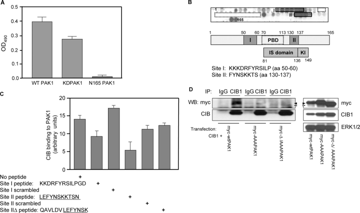
Figure 2.
Identification of CIB1-binding sites within the PAK1 NH 2 terminus. (A) CIB1 binds to full-length wild-type GST-PAK1 (wtPAK1) and GST-PAK1 K298A (kdPAK1) but not the NH2-terminal deletion GST-N165-PAK1. Solid-phase binding assays were performed as in Fig. 1 C, and soluble GST-PAK1, GST-PAK1 K298A, and GST-N165-PAK1 were added to wells coated with CIB1. (B) Delineation of the CIB1-binding sites in the PAK1 NH2-terminal sequence by SPOT peptide method. Top and middle panels show two sets of CIB1 reactive spots labeled as sites I and II relative to the PBD- or Rac/Cdc42-binding sites (residues 70–113, dotted boxes). The bottom panel indicates the inhibitory switch (IS) and kinase inhibitor (KI) domains. Sequences of corresponding CIB1-binding sites are below. (C) Inhibition of CIB1 binding to PAK1 with site I and II peptides. Ni-NTA agarose beads loaded with purified His-PAK1 were added to recombinant CIB1 that was preincubated with and without PAK1 peptides corresponding to sites I and II and scrambled sites I and II. Peptide sequences of sites I, II, and IIΔ are shown below. The graph represents densitometry of affinity precipitates probed for CIB1 and normalized to His-PAK1 that was immunoblotted from the same membrane. Data represent SEM from two independent experiments (error bars). (D) Loss of CIB1 binding to site I and II PAK1 mutants. Clarified lysates from HEK293 cells cotransfected with CIB1 and myc wild-type (wt)PAK1, myc-PAK1 siteIAAA (myc-AAAPAK1), or myc-PAK1 siteIΔ/siteIIAAA (myc-Δ/AAA PAK1) were immunoprecipitated with a control IgG or anti-CIB1 antibody and with samples immunoblotted for myc (top) and CIB1 (bottom; n ≥ 4). Western blots of the input expression of CIB1 and myc-tagged wtPAK1 (closed arrowheads), AAAPAK (closed arrowheads), or Δ/AAAPAK1 (open arrowheads).

