Figure 7.
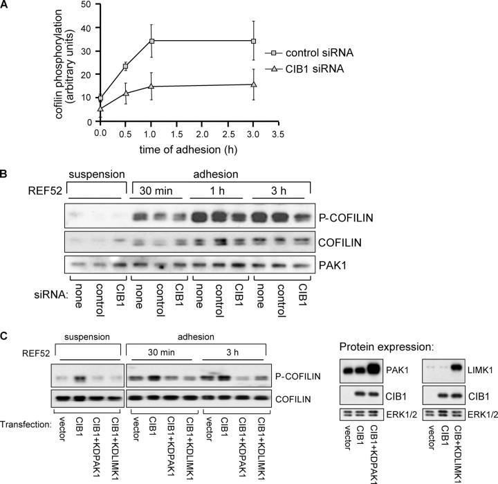
CIB1 modulates downstream signaling to cofilin. (A) Lysates were prepared from control or specific CIB1 siRNA-transfected REF52 cells either held in suspension or adhered to FN for the indicated times. Densitometry of p-cofilin levels from lysates that were prepared from control or specific CIB siRNA was normalized to ERK or PAK1 from the same blots. Error bars represent means ± SEM (n = 2). (B) Representative membrane immunoblotted with antibodies against total or phosphorylated cofilin (p-cofilin). The top half of the membrane was also immunoblotted for PAK1 expression (bottom). (C) Lysates prepared from REF52 cells overexpressing empty vector or CIB1 ± kdPAK1 or kdLIMK1 were analyzed for cofilin phosphorylation as in A. The membrane was reprobed for total cofilin (bottom). Lysates from cells expressing control vector, CIB1, or CIB1 coexpressed with kdPAK1 or kdLIMK1 were immunoblotted using anti-CIB1, -PAK1, or -LIMK1 antibodies (middle and right). PAK1 immunoblots show both endogenous PAK1 and overexpressed kdPAK1 (top middle). Immunoblotting for LIMK1 also shows endogenous LIMK1 and overexpressed kdLIMK1 (top right). Middle blots show overexpressed CIB1. Membranes were also probed with an anti-ERK antibody as a loading control (bottom, middle and right). Data represent two separate experiments.

