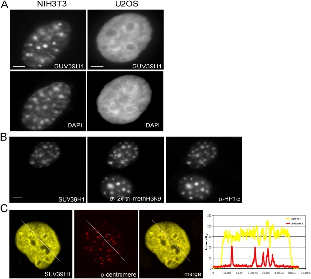
Figure 1.
Localization of EYFP-SUV39H1 in mouse NIH3T3 as well as in human U2OS cells. (A) After transfection, EYFP-SUV39H1 localizes in NIH3T3 cells to distinct nuclear regions that are also visible by DAPI staining. In U2OS cells, EYFP-SUV39H1 also localizes at sites that are stained by DAPI, but these areas are less well defined. (B) NIH3T3 cells transfected with EYFP-SUV39H1 were labeled with antibodies specific for trimethylated H3K9 and HP1α. (C) Human U2OS cells transfected with EYFP-SUV39H1 were labeled with antibodies against centromeres. Single optical sections show the YFP-tagged protein, centromere labeling, and an overlay. Line scans (diagonal lines through the images) show the local intensity distributions of the EYFP fusion protein in yellow and of the centromere labeling in red. Bars, 10 μm.

