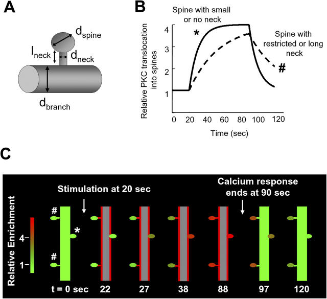
Figure 8.
A model of the spine translocation process shows that spine necks provide a means to suppress subthreshold stimuli, integrate activity, and create local memory of past activity. (A) Diagram illustrating the parameters used to simulate translocation of PKCγ into dendritic spines. (B) Simulation of PKCγ translocation curves for two spines with different morphologies. Translocation was compared between a spine that only has a small neck or no neck at all and one that has a restricted neck. The spine with the small neck (*) is a fast-loading synapse and filled rapidly with PKC, unlike the spine with the restriction (#), which loaded much more slowly. (C) Schematic images were drawn by a MatLab program based on simulations of PKCγ translocation from a main branch into three different synaptic spines. One spine had no neck constriction, and two spines had neck constrictions because of long or narrow necks (see Materials and methods for model descriptions). Translocation into the spine without a neck was rapid and reversible, whereas the spines with restricted necks underwent a slow PKCγ recruitment behavior, and the protein remained in the spine for a much longer time than the spine with no neck. Red indicates a high concentration of PKCγ at the plasma membrane or in spines, whereas green shows a low concentration of PKCγ.

