
Figure 3.
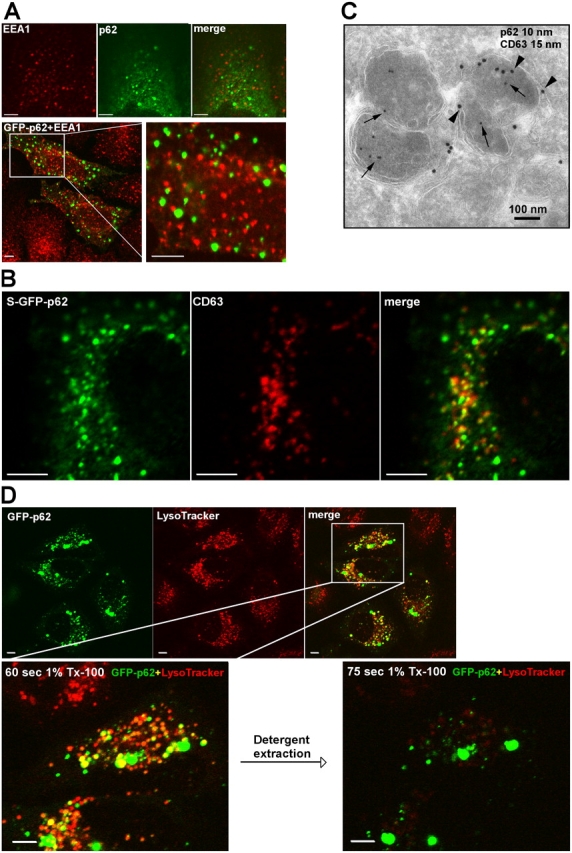
p62 bodies are found both as membrane-free protein aggregates and as membrane-confined autophagosomal and lysosomal structures. (A) Localization of bodies containing endogenous p62 or transiently expressed GFP-p62 relative to EEA1-positive early endosomes. Endogenous p62 were stained green using p62 antibodies directly labeled with AlexaFluor488, and EEA1 was stained red with EEA1 mAbs directly labeled with AlexaFluor555. Alternatively, transiently expressed GFP-p62 was expressed in HeLa cells, and EEA1 was stained red with EEA1 mAb (bottom). The boxed area is shown to the right at a higher magnification. (B) Colocalization of GFP-p62 and CD63 (stained red using a mAb) in HeLa cells stably expressing GFP-p62. (C) Immunoelectron micrograph of S–GFP-p62 stained with a GFP pAb (10-nm gold particle, arrows) and monoclonal CD63 (15-nm gold particle, arrowheads). (D) Rapid detergent extraction of GFP-p62 from LysoTracker-positive acidic organelles. HeLa cells transiently expressing GFP-p62 were labeled with LysoTracker for 60 min. The detergent extractions were imaged in a time series with 15-s time intervals after adding 1% Triton X-100. The boxed area is shown in the bottom two images at a higher magnification before and after detergent extraction. Bars (A, B, and D), 20 μm.