Figure 4.
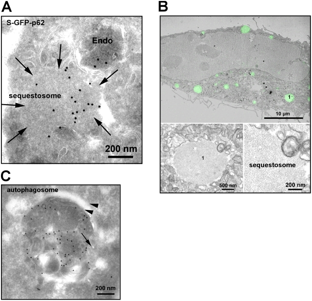
p62 is found both in autophagosomes and in cytosolic aggregates/sequestosomes. (A) Immuno-EM of GFP-p62. S–GFP-p62 cells were labeled with rabbit anti-GFP (Abcam) followed by protein A–gold (15 nm). We observed labeling in membrane-free cytosolic structures and sequestosomes (arrows) as well as in endosomes (Endo). (B) Correlative immunofluorescence/EM of HeLa cells treated with 10 μM PSI for 5 h displaying typical sequestosomes. The insets show two magnifications of the sequestosome, which is labeled 1. (C) Representative image of a p62-containing autophagosome. HeLa cells transfected with GFP-p62 were immunogold labeled as in A. Note the cisternal-like membrane (arrowheads) surrounding the GFP-positive material. The arrow indicates a fused endosome.

