
Figure 3.
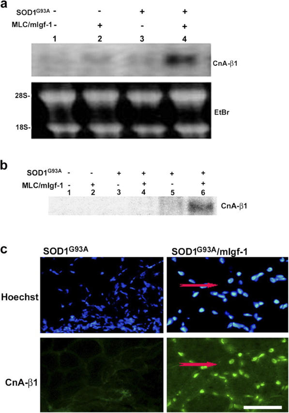
Transgenic mIgf-1 expression induces chronic CnA-β1 expression in SOD1G93A mice. (a) Northern blot analysis of CnA-β1 expression in wt (lane 1), MLC/mIgf-1 (lane 2), SOD1G93A (lane 3) and SOD1G93A/mIgf-1 (lane 4) transgenic mice. Ethidium bromide staining was used to verify equal loading of the RNA sample. (b) Lysates of the same muscle tissues used in Fig. 2 c were tested by Western blotting using a CnA-β1–specific antibody. (c) Immunofluorescence of transverse sections from quadriceps muscles of SOD1G93A and SODG93A/mIgf-1 at paralysis stage. CnA-β1 shows a nuclear localization. A regenerating fiber is indicated by the presence of central nucleus (red arrows). Nuclei were visualized by Hoechst dye (blue). Bar, 20 μm.