Figure 1.
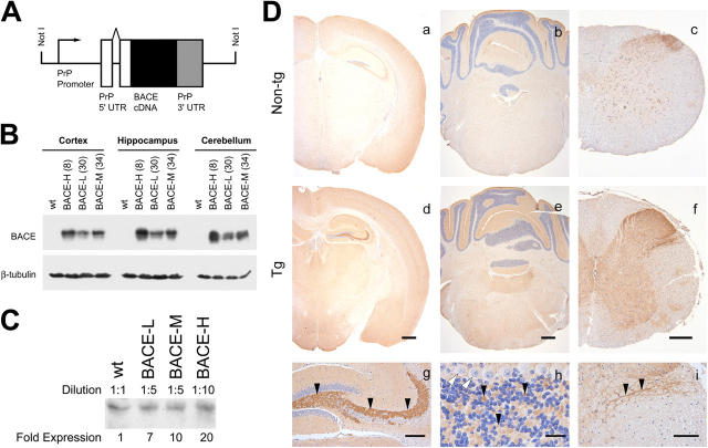
Generation of Tg mice overexpressing human BACE. (A) Schematic of the expression cassette used to generate PrP-BACE mice, containing the human BACE cDNA flanked by the 5′ UTR of the murine PrP gene containing an intron, and the 3′ UTR of the PrP gene. (B) BACE expression in Tg mice. RIPA lysates from the cortex, hippocampus, and cerebellum were immunoblotted for BACE using an antibody against the COOH terminus of human BACE. Three Tg lines (n > 3; shown are mice at 14–16 mo) were analyzed with low (30), medium (34), and high (8) expression. (C) BACE expression relative to non-Tg mice. Cortical RIPA lysates were diluted as indicated before immunoblotting for BACE using a polyclonal antisera against the entire NH2-terminal ectodomain of BACE. BACE-L, -M, and -H mice overexpress ∼7-, 10-, and 20-fold more BACE than non-Tg mice (n > 3 per genotype). No age-dependent changes in BACE expression have been observed (up to 20 mo). (D) Regional expression of BACE. Immunohistochemistry using BACE-N1 demonstrates increased immunoreactivity in BACE overexpressing mice. Representative images are shown of the cortex, striatum, cerebellum, brainstem, and spinal cord. Higher magnification shows staining of mossy fiber terminals, the granule cell layer of the cerebellum, and the dorsal spinal cord. Sections from APP (a–c) and BACE-H (d–i) mice are shown. Black arrowheads demonstrate synaptic/axonal staining, whereas white arrowheads show Purkinje cells. Wild-type/APP or BACE/BACExAPP mice showed no differences in staining (n > 2 per genotype). Similar staining was observed with a polyclonal antibody raised against the COOH terminus of BACE and polyclonal antibodies raised against the ectodomain of BACE. Bars: (a and d) 500 μm; (b and e) 500 μm; (c and f) 250 μm; (g) 150 μm; (h) 25 μm; (i)100 μm.

