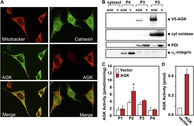
Figure 2.
Subcellular localization of AGK. (A) NIH 3T3 fibroblasts were transiently transfected with V5-tagged AGK and mitochondria stained with MitoTracker red. The ER was visualized with anti-calnexin antibody followed by FITC-conjugated anti-rabbit as the secondary antibody. AGK was stained with monoclonal anti-V5 antibody followed by secondary FITC-conjugated or Texas red–conjugated anti–mouse antibody. Cells were visualized by dual wavelength confocal microscopy. Superimposed merged pictures are shown in the bottom panels, with yellow indicating colocalization. (B and C) Activity and expression of AGK in subcellular fractions. Lysates from HEK 293 cells transfected with vector or V5-AGK and P2 (mitochondria), P3 (ER and Golgi), P4 (plasma membrane), and cytosol fractions isolated. The P1 fraction containing nuclei and unbroken cells was not examined. 25 μg of proteins were resolved by SDS-PAGE and immunoblotted with anti-V5 antibody or with antibodies to the specific organelle markers anti–cytochrome c oxidase, anti-phosphodisulfide isomerase (PDI), and anti–αv-integrin. AGK activity was also determined in each subcellular fraction with MOG as substrate. Results are means ± SD of triplicate determinations. Similar results were obtained in two additional experiments. *, P < 0.05 by t test. (D) 400-μg aliquots of lysates from HEK 293 cells transiently transfected with vector (open bars) or V5-AGK (closed bars) were immunoprecipitated with anti-V5 antibody as described in Materials and methods, and AGK activity was determined in the immunoprecipitates. Data are expressed as picomoles of LPA formed in 30 min and are means ± SD of duplicate determinations.

