Figure 6.
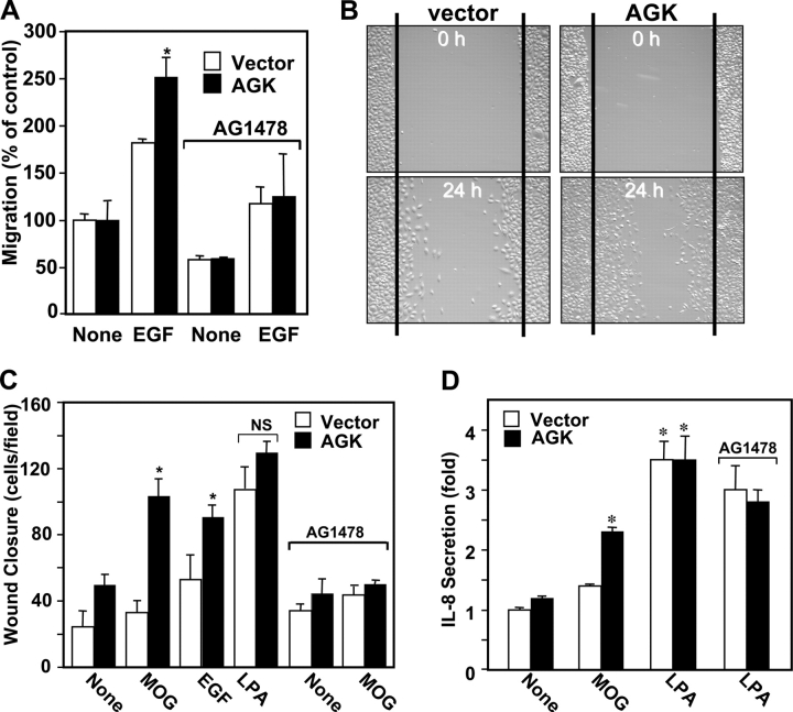
EGFR is required for AGK-stimulated cell migration toward EGF and wound closure. (A) PC-3 cells transfected with vector (open bars) or AGK (closed bars) were pretreated without or with 200 nM AG1478 for 20 min and allowed to migrate for 3 h toward EGF (10 ng/ml). The data are means ± SD of two determinations. Similar results were obtained in two independent experiments. (B and C) Monolayers of vector (open bars) or AGK (closed bars) PC-3 transfectants were wounded and treated with vehicle, MOG (10 μM), LPA (10 μM), or EGF (10 ng/ml). Where indicated, cells were also treated with 200 nM AG1478. (B) Representative images of a wound healing assay with vector and AGK-transfected PC-3 cells before and 24 h after treatment with MOG. (C) Migration of cells into the wound was determined after 24 h by processing digital photographs with ImagePro Plus. (D) AGK induces IL-8 secretion. PC-3 cells transfected with vector (open bars) or AGK (closed bars) were serum starved for 24 h and treated in serum-free DME with or without MOG (10 μM) or LPA (1 μM) for 16 h, and IL-8 secretion was measured by ELISA. Where indicated, cells were also treated with 200 nM AG1478. *, P < 0.05 by t test.

