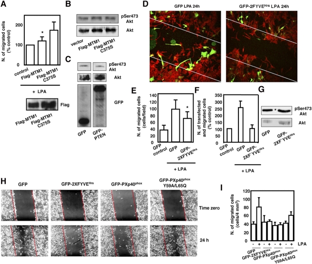
Figure 6.
PtdIns-3-P is involved in the LPA-mediated migration of SKOV-3. (A-G) SKOV-3 were transiently transfected with the indicated cDNAs. Transfection efficiency in these cells was ∼50%. (A, top) SKOV-3 were transfected with cDNAs encoding Flag-MTM1 and Flag-MTM1 C375S. After 24 h, cells were serum deprived overnight and migration was assessed by Transwell assay in the presence of 25 μM LPA. The total number of migrated cells was counted after staining the membranes of the Transwell chambers with crystal violet. Data are mean ± SEM of three independent experiments. *, P < 0.05. (bottom) Representative blot monitoring the levels of expression of Flag-MTM1 and Flag-MTM1 C375S by using an anti-Flag antibody. (B) Phosphorylation of Akt on residue Ser473 assessed in lysates from SKOV-3 transfected with an empty vector or cDNAs encoding Flag-MTM1 and Flag-MTM1 C375S. Filters were stripped and reprobed with an anti-Akt antibody. (C) Phosphorylation of Akt on residue Ser473 assessed in lysates from SKOV-3 transfected with cDNAs encoding GFP or GFP-PTEN. Filters were stripped and reprobed with an anti-Akt antibody. Expression of the exogenous proteins was assessed by using an anti-GFP antibody. Images are from different lanes in the same blot. (D) SKOV-3 grown on glass coverslips were transfected with cDNAs encoding GFP or GFP-2XFYVEHrs. After 24 h, cells were serum deprived overnight and migration was assessed by wound healing assay. After wounding and further 24 h in the presence of 25 μM LPA, cells were fixed and analyzed by confocal microscopy. Actin was stained by using Alexa 594 phalloidin and the merged images are shown. Diagonal lines specify the position of the original wound. (E and F) SKOV-3 were transfected as in D. Migration was then assessed by Transwell assay in the presence of 25 μM LPA. For data shown in E, the total number of cells that had migrated was counted after staining the membranes of the Transwell chambers with crystal violet. Data are mean ± SEM from three independent experiments performed in duplicate. *, P < 0.05. For data shown in F, the membranes of the Transwell chambers were fixed and analyzed by fluorescent microscopy to visualize and count only transfected (green) cells that had migrated. Data are mean ± SEM from four independent experiments. (G) Phosphorylation of Akt on residue Ser473 assessed in lysates from SKOV-3 transfected with cDNAs encoding GFP or GFP-2XFYVEHrs. Filters were stripped and reprobed with an anti-Akt antibody. (H–I) HeLa were transfected with cDNAs encoding the indicated fusion proteins (transfection efficiency was >80%). After 24 h, cells were serum deprived overnight and wound healing was performed in the presence of 25 μM LPA. (H) Representative phase-contrast images of the wounded cell monolayers at time 0 and after 24 h of migration. (I) Quantitative analysis of wound healing as in H. Data are mean ± SEM of three independent experiments.

