
Figure 9.
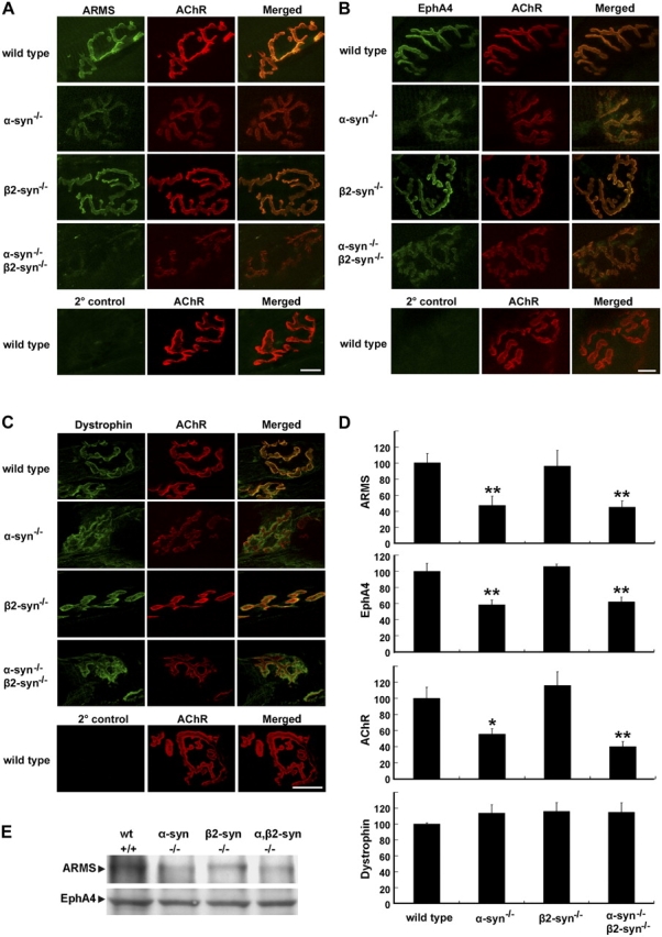
Abnormal synaptic localization of ARMS and EphA4 in syntrophin-null mice. (A) Longitudinal sections of sternomastoid muscle were double stained with α-BTX and ARMS antibody (A), EphA4 antibody (B), or dystrophin antibody (C). Wild-type sections were stained with FITC-conjugated secondary antibody and α-BTX to show the background levels. Bars (A and B), 10 μm; (C), 20 μm. (D) Quantitative analysis showed reduced junctional staining of ARMS and EphA4 in α- and α,β2-syntrophin–null mice, but not for dystrophin. y axis indicated the knockout NMJ compared with that in the wild-type NMJ, which was normalized to 100%. n = 5; *, P < 0.05; **, P < 0.01. Error bars represent the SEM. (E) Membrane proteins of gastronemius muscle from syntrophin-null mice and wild-type littermates were prepared and subjected to Western blot with ARMS and EphA4 antibodies.