
Figure 1.
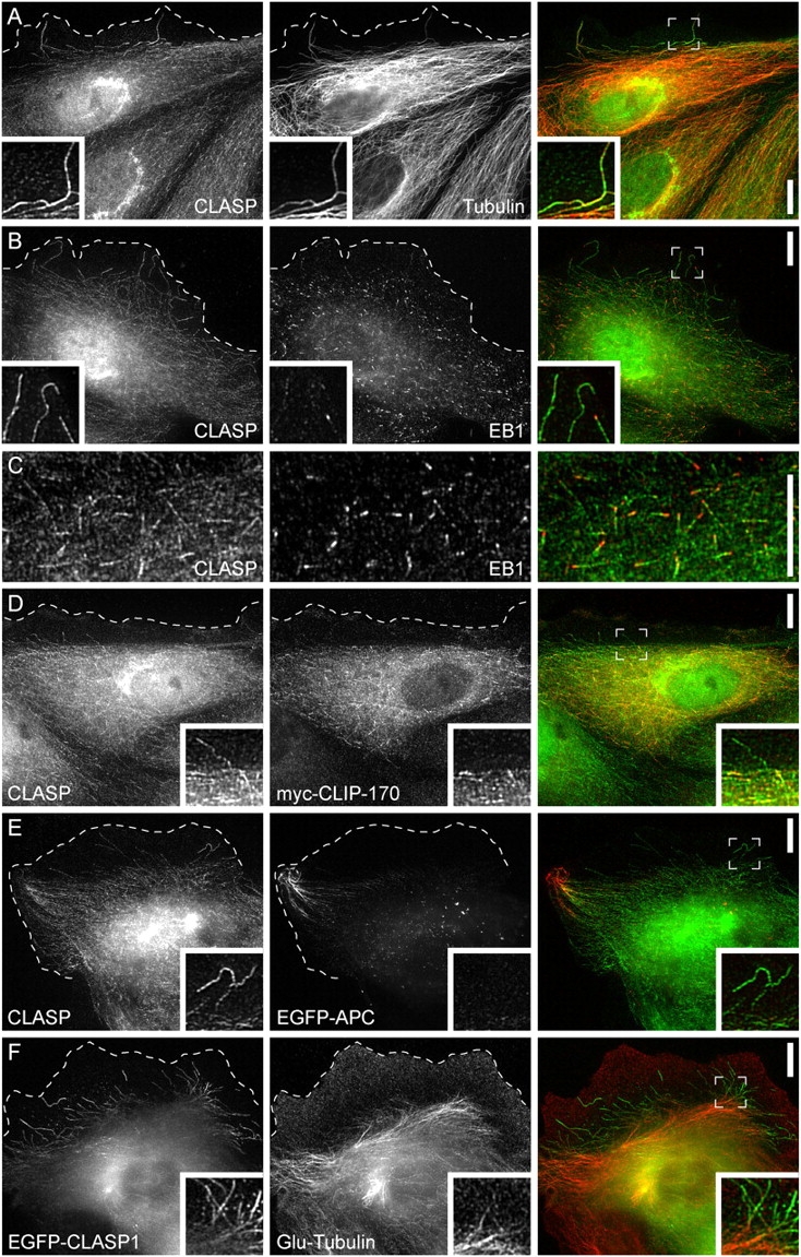
CLASPs are localized along MT lattices in the lamella and to MT plus ends in the cell body. (A) Immunofluorescence of CLASPs and MTs in PtK1 epithelial cells. Cells were stained with antibodies against the Xenopus CLASP homologue and MTs. (B) Immunofluorescence of CLASPs and EB1. (C) Higher magnification of CLASP- and EB1-MT plus end association in the cell body. EB1 is most concentrated at the very tip of the MT, whereas CLASPs are localized slightly behind EB1. (D) Immunofluorescence of CLASPs and expressed myc-tagged CLIP-170. (E) Immunofluorescence of CLASPs and expressed EGFP-tagged APC. None of the other +TIPs show specific accumulation on the lattice of lamella MTs. (F) Distribution of detyrosinated tubulin in a cell expressing EGFP-tagged CLASP1. Insets show lamella cell regions at higher magnification and the dashed line indicates the protruding cell edge. Bars, 10 μm.