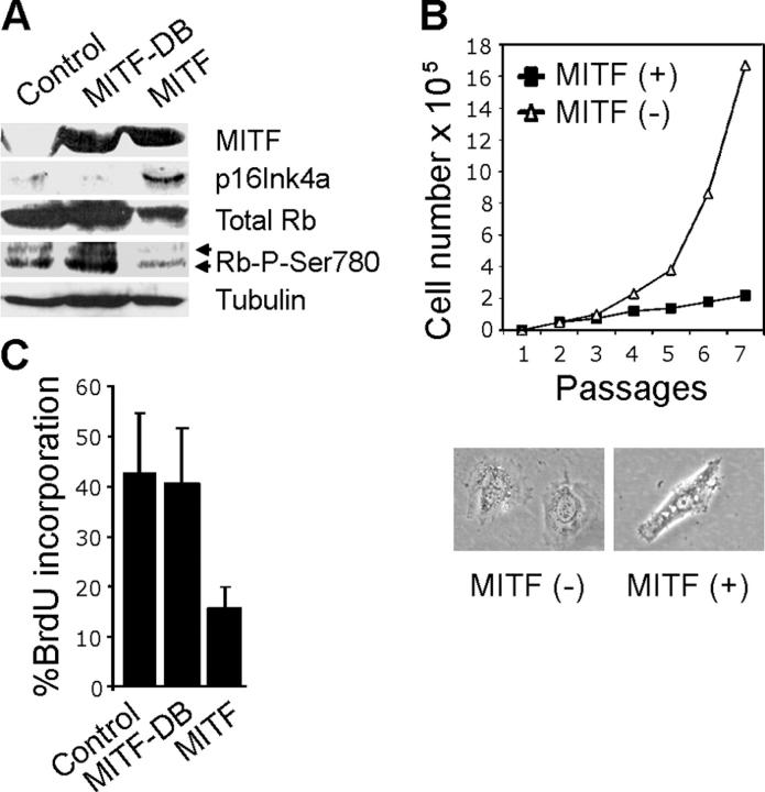
Figure 1.
MITF inhibits cell proliferation. (A) Western blot demonstrating MITF, p16Ink4a, total Rb, Rb-phospho-Ser780, and tubulin protein expression in 10T1/2 mouse fibroblasts stably expressing ectopic MITF, MITF-DB (DNA-binding mutant), or empty control vector. Arrows indicate hyperphosphorylated (top band) and hypophosphorylated (bottom band) forms of Rb. (B) Growth curves of 10T1/2 cells stably expressing MITF or empty control vector. Panels show morphology of 10T1/2 cells expressing control empty vector (left) and MITF (right). (C) BrdU incorporation assays in 10T1/2 cells expressing ectopic MITF , MITF-DB, or empty control vector.

