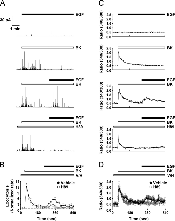
Figure 1.
Sensitization of the functionally silent EGF-induced [Ca2 + ]i increase and exocytosis by BK in rat adrenal chromaffin cells. (A) Carbon fiber amperometry was used to detect exocytosis from single chromaffin cells in real time. Typical amperometric responses from adrenal chromaffin cells after addition of 20 nM EGF and/or 1 μM BK are shown. Involvement of PKA was examined by application of H89 (10 μM) before BK stimulation. (B) Normalized rate of exocytosis. Number of amperometric spikes over a 20-s recording period was divided by that over a 20-s control period. Each point represents a mean ± SEM value obtained from 15 control and 12 H89-treated cells from three separate experiments on different batches of cells. The normalized rate of exocytosis in the control period is assigned a value of 1.0 and indicated by a horizontal broken line. (C and D) Fura-2 intracellular Ca2+ imaging of chromaffin cells in response to 1 μM BK and/or 20 nM EGF stimulation. H89 (10 μM) was treated before BK stimulation as indicated. Representative Ca2+ transients are shown in C, and averaged Ca2+ traces from multiple cells are shown in D. At least three independent experiments were performed and cells from two coverslips were analyzed per experiment.

