Figure 2.
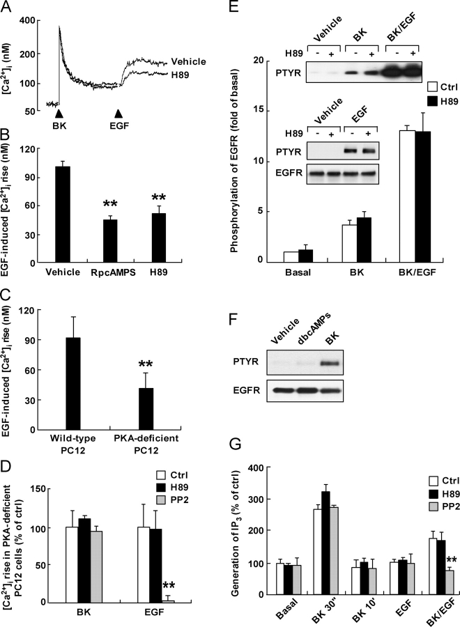
Involvement of PKA in the sensitization process. (A) Fura-2–loaded PC12 cells (106) were pre-treated with either vehicle or H89 (10 μM), followed by BK, and then finally stimulated with EGF. Representative traces from more than three independent experiments are shown. (B) Fura-2–loaded PC12 cells were pre-treated with H89 (10 μM) or RpcAMPS (100 μM), followed by BK, and then finally stimulated with EGF. (C) EGF-induced [Ca2+]i increase subsequent to BK stimulation were compared in fura-2–loaded wild-type and PKA-deficient PC12 cells. (D) PKA-deficient cells were pretreated with vehicle, H89 (10 μM), or PP2 (10 μM), followed by BK, and then finally stimulated with EGF. (B–D) Peak amplitudes in the BK- and/or EGF-induced [Ca2+]i rise were measured. (E) PC12 cells were treated with vehicle, BK (2 min), EGF (200 pM, 2 min), or sequentially with BK (10 min) followed by EGF (200 pM, 2 min) in the presence or absence of H89 (10 μM) as indicated. Cell lysates were immunoprecipitated with the EGFR antibody, and the precipitates were immunoblotted with anti-phosphotyrosine antibody. Immunoblotting with the EGFR antibody was performed for normalization. EGFR phosphorylation was quantitated by densitometry and normalized. (F) PC12 cells were treated with vehicle, dibutyryl cAMP (1 mM), or BK for 3 min and tyrosine phosphorylated EGFRs were monitored as in E. (G) PC12 cells were stimulated as indicated and the level of IP3 was measured. BK/EGF denotes for cells stimulated with EGF (1 min) after BK (10 min) pretreatment. H89 (10 μM) or PP2 (10 μM) was pretreated for 3 min. The concentration of BK was 1 μM and EGF was 20 nM, unless otherwise stated. All error bars are mean ± SEM of a minimum of three experiments. **, P < 0.01.

