Figure 4.

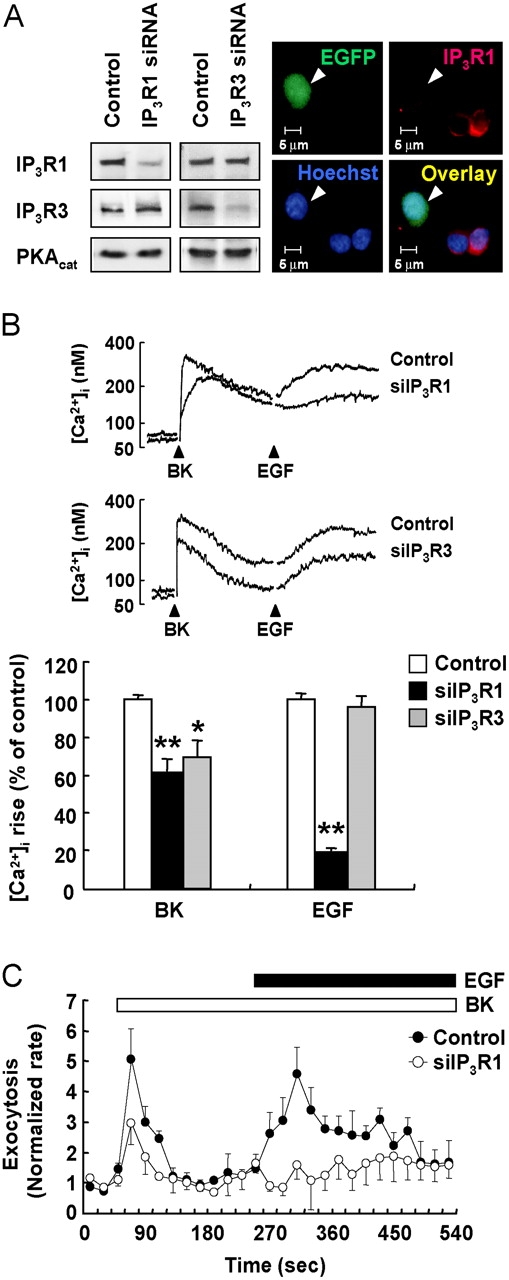
IP3R1 is required in the sensitization process. (A) PC12 cells were transfected with siRNA against IP3R1 or IP3R3 as indicated. pEGFP was cotransfected as a marker. Effects of siRNA were confirmed by Western blot analysis and immunocytochemistry. (Left) Representative immunoblots after transfection with siRNA against IP3R1 or IP3R3. (Right) Typical images after transfection with IP3R1 siRNA. (B) Cells transfected with siRNA as indicated were loaded with fura-2 for Ca2+ measurements. Cells were treated with 1 μM BK, followed by 20 nM EGF. Typical Ca2+ transients are presented, and the peak heights in the BK- and EGF-induced [Ca2+]i increase were measured. *, P < 0.05; **, P < 0.01. (C) Carbon fiber amperometry was used to detect exocytosis from single cells in real time. Control or IP3R1 siRNA-transfected PC12 cells were treated with 1 μM BK followed by 20 nM EGF as indicated. The rate of exocytosis was normalized as in Fig. 1 B. Each point presented is a mean ± SEM of three experiments.
