
Figure 4.
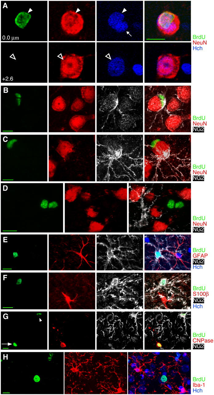
Many cells in the adult rat cortex do not appear to be neurons or commonly recognized glial types. (A) BrdU+ satellite cells closely apposed to large neurons could lead to erroneous identification of the large neurons as BrdU labeled. However, Hoechst nuclear counterstain and z-sectioning make it clear that the 4–5-wk-old BrdU+ cell nucleus (arrowhead at 0.0 μm) is not the nucleus of the large neuron (hollow arrow at 2.6 μm) and must instead belong to a satellite cell. A second, BrdU−, satellite cell can also be seen with Hoechst counterstain (small arrow at 0.0 μm). (B and C) Some but not all 4–5-wk-old BrdU+ satellite cells express the chondroitin sulfate proteoglycan NG2, characteristic of dividing or immature cells. Two satellite cells, one BrdU+/NG2− and one BrdU−/NG2+ can be seen in C. (D) A pair of 4–5-wk-old BrdU+ cells are NG2− and NeuN−, as were ∼40% of 4–5-wk-old BrdU+ cells. (E–G) Newborn NG2+ cells do not stain for astrocyte, oligodendrocyte, or microglial markers. (E) A BrdU+/NG2+ cell and a BrdU−/GFAP+ cell 2 h after BrdU injection. (F) A BrdU+/NG2+ cell and a BrdU−/S100β+ cell 2 h after BrdU injection. (G) A 3–4-wk-old BrdU+ cell in the cortex expressing the oligodendrocyte marker CNPase (arrow) shows faint nuclear staining of NG2 around the nucleus but none of the NG2+ processes that defined NG2+ cells. A second BrdU+ cell (arrowhead) shows strong NG2 staining and very faint CNPase staining. (H) A solidly stained BrdU+ cell and a pair of cells with speckled BrdU staining, all 4–5 wk old and all nonimmunoreactive for the microglial marker Iba-1. Images show single planes (B and E) or Z-axis projections of 10 × 0.69 μm (C), 7 × 1.05 μm (D), 8 × 1.72 μm (F), 9 × 0.67 μm (G), or 18 × 1.38 μm (H). Bars: 5 μm in orthogonal views; 10 μm in all other views.