
Figure 2.
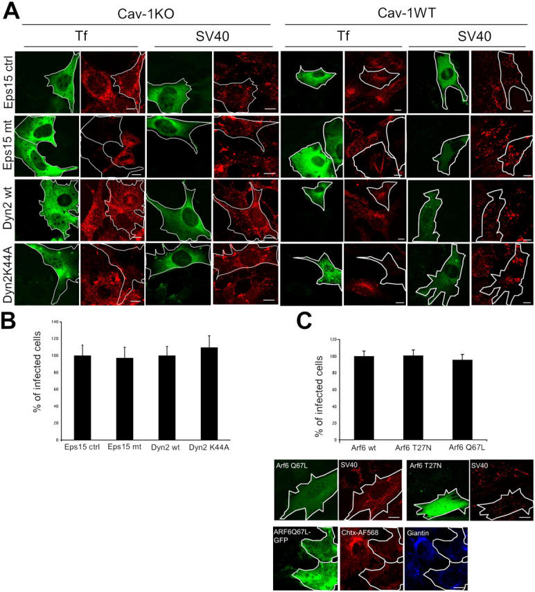
Internalization and infection of SV40 occurs independently of Eps15, Dynamin II, and ARF6. Cav-1KO and cav-1WT cells were transfected with Eps15IIIΔ2 (Eps15 ctrl), Eps15 EΔ95/295 (Eps15 mt), Dyn2wt, or Dyn2K44A, all tagged with GFP. (A) After transfection, cells were incubated with AF594-SV40 for 2 h, fixed, and examined in confocal microscopy. AF594-Tf served as a positive control. Representative images are shown. Bars, 10 μm. (B) After transfection, cav-1KO cells were infected with SV40 for 20 h, fixed, and analyzed for infection. Infection in cells expressing the control constructs was set at 100%. Values are given as the mean ± SD. (C) Cav-1KO cells were cotransfected with ARF6 wild type, ARF6 T27N, ARF6 Q67L, and GFP. After transfection, cells were infected with SV40 for 20 h, fixed, and analyzed for infection. Infection in cells expressing the wild-type construct was set at 100%. Values are given as the mean ± SD. Alternatively, cells were incubated with AF594-SV40 for 1.5 h and imaged live. As a positive control, ARF6 Q67L-GFP–transfected cells were incubated with cholera toxin-AF568, fixed, and immunostained with an anti-giantin antibody (blue). Bars, 10 μm.