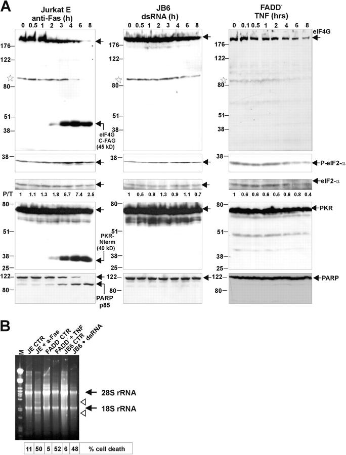
Figure 3.
Translation initiation factors eIF4G, eIF2-α, PKR, and PARP remain intact in necrosis but not in apoptosis. (A) Western blot analysis of total cell lysates reveals the cleavage of eIF4G into a 45-kD fragment (eIF4G C-FAG) in time after anti-Fas treatment of JE cells (left). Asterisks denote an aspecific band. The phosphorylation status of eIF2-α was monitored using an antibody specific for eIF2-α phosphorylated at Ser51 (P-eIF2-α). The total amount of eIF2-α was also assessed on the same blot after inactivation of the P-eIF2-α signal by overnight treatment of the blot with 0.1% NaN3. The ratio of phosphorylated over total eIF2-α signal per lane was determined by densitometric analysis, normalized to 1 for time 0, and is indicated below each lane. Immunoblots for PKR and PARP show the appearance of a 40-kD PKR fragment (PKR-Nterm) and of an 85-kD fragment of PARP in apoptosis. (B) Ribosomal 28S RNA is cleaved in apoptosis but not in necrosis. 10 μg of total RNA isolated from cells treated as indicated was analyzed by agarose gel electrophoresis followed by ethidium bromide staining. Arrows indicate the position of 28S and 18S rRNA. Arrowheads indicate cleavage products in the anti-Fas–treated sample. CTR, untreated.

