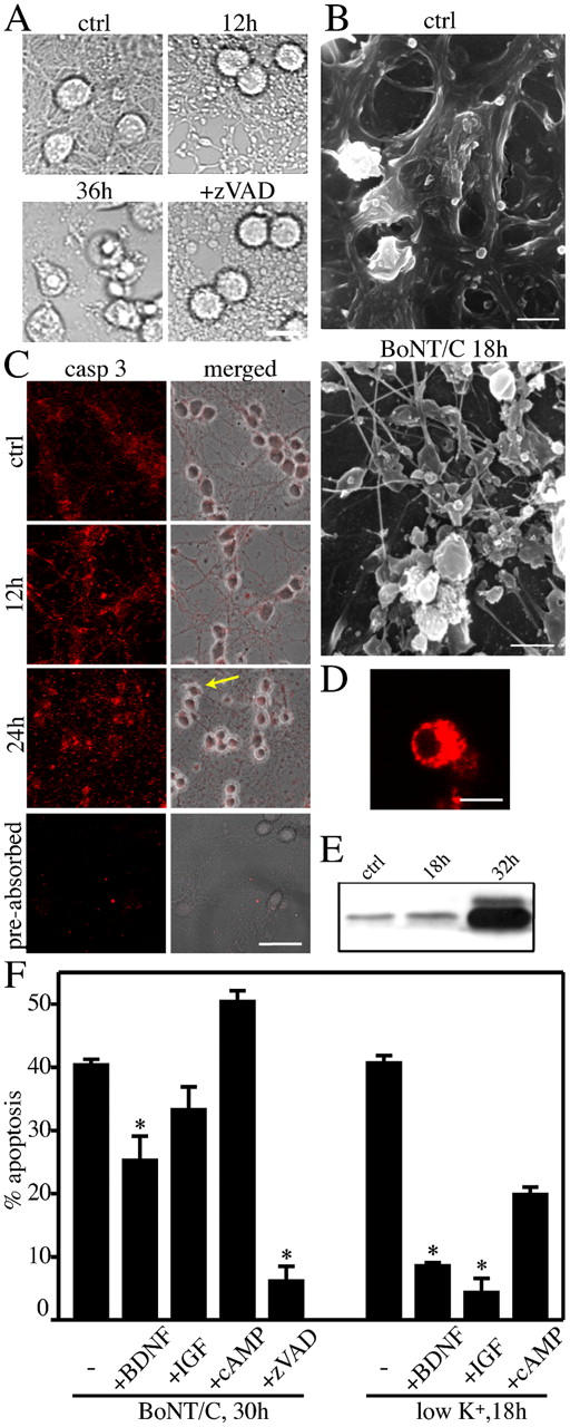
Figure 4.

A caspase-independent axonal breakdown preceded the apoptotic demise of the neuronal bodies. Appearance of the axo-dendritic network in CGC cultures as shown by phase-contrast (A) and scanning electron microscopy (B). The disassembly of the neuronal projections in the presence of 20 ng/ml BoNT/C is noteworthy. The magnification used in B allows the visualization of the only neuronal network. The p17 fragment of caspase-3 was detected along the neuronal projections (C), as well as within the cell bodies (D), by confocal microscopy. Note the absence of staining when the antibody was preabsorbed with p20/p17 caspase-3. Fluorescence images in C represent single focal planes acquired at the level of the neurites. The arrow indicates the cell shown in D to illustrate the presence of processed caspase-3 at the level of the cell body at 24 h. The Western blot shows small amounts of caspase-3 processing in untreated neurons and 18 h after BoNT/C. Substantial activation is seen after 32 h, when most neurons have undergone apoptosis (E). Note that caspase inhibition by zVAD-fmk prevented nuclear condensation, but did not prevent the dismantling of the neurites (A). Neurotrophic factors (100 ng/ml BDNF and 200 ng/ml IGF-1) and cAMP (1 mM) did not prevent axonal degeneration (not depicted) and only BDNF partially protected from apoptosis (E). Neurotrophic factors efficacy was monitored in parallel cultures in which apoptosis was induced by K+ withdrawal. The percentage of apoptosis was evaluated by scoring condensed, H33342-positive nuclei. (F) Data are means ± SD for triplicate determinations; *, P < 0.005. Bars: (A) 10 μm; (B) 5 μm; (C) 30 μm; (D) 10 μm.
