Figure 5.

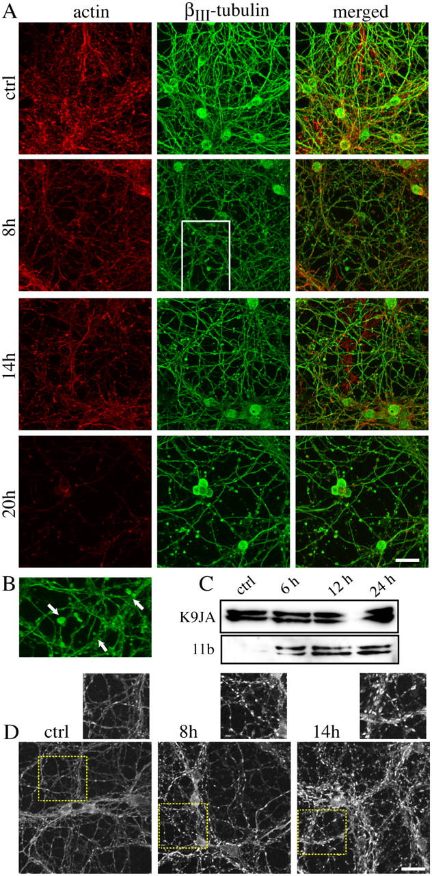
Cytoskeletal disassembly and abnormal tau phosphorylation accompanied the early degeneration of the neurites. (A) CGCs were exposed to 20 ng/ml BoNT/C for the times indicated and immunostained for actin and βIII-tubulin. Confocal images were acquired from the same field. The bulblike structures present at 8 h along the neurites are shown at higher magnification in B (arrows). (C) The total amount of tau protein was detected by the K9JA antibody, which recognizes tau independently of its phosphorylation. In the same cell extracts, abnormal tau phosphorylation was detected by using the S202/205 phosphospecific antibody 11b. Neurons were exposed to 20 ng/ml BoNT/C for the indicated times. (D) The pattern of tau distribution was determined by a different antibody (Innogenetics) and changed in cells exposed to 20 ng/ml BoNT/C. Images in A, B, and D are extended focus images derived by merging 50–60 focal planes acquired in the z dimension with a 63× oil 1.3 NA apochromat objective. Bars, 20 μm.
