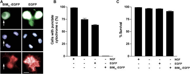
Figure 5.
BIMEL expression promotes loss of mitochondrial cytochrome c but does not decrease survival in NGF-deprived neurons exposed to low O2. Neurons were infected with Ad-BIMEL/EGFP or Ad-EGFP adenoviruses (MOI = 100). The next day, NGF was withdrawn and the cells were immediately transferred to 1% O2. (A and B) After 24 h of NGF deprivation, the cells were fixed and analyzed for immunofluorescence using anti–cytochrome c antibody. The three panels for each treatment are of the same field of view. Bar, 15 μm. The percentage of EGFP-expressing, BIMEL/EGFP-expressing, and control (uninfected) cells showing punctate cytochrome c immunofluorescence was determined (means ± SEM, n = 3). Arrows in A point to a neuron expressing BIMEL-EGFP (top), its Hoechst-stained nucleus (middle), and its lack of punctate cytochrome c immunofluorescence (bottom). (C) Neurons were treated as described above except that NGF deprivation was continued for 48 h. The cells were then stained with Hoechst dye and scored for viability (mean ± SEM, n = 3). Except in the case of uninfected cells, only neurons positive for BIMEL/EGFP or EGFP expression were scored in B and C.

