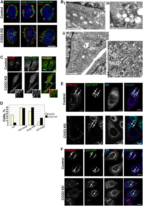
Figure 8.

Golgi in COG3 KD cells is disrupted to mini-stacks, which are proficient in anterograde VSVG delivery to plasma membrane and defective in retrograde trafficking of Shiga toxin B subunit. (A) Control and COG3 KD cells that stably express GalNAcT2-GFP were fixed and stained with anti-p115, anti-giantin, or anti-p230 antibodies and secondary antibodies conjugated with Alexa 594. DNA was stained with DAPI. Images were acquired with 100× objective and deconvolved. Bar, 10 μm. (B) Ultrastructural analysis of Golgi in COG3 KD cells. Electron micrographs of the juxtanuclear region in control (i) and COG3 KD (ii and iii) cells. Note the Golgi mini-stack in ii and multiple 60-nm vesicles in COG3 KD cells (ii and iii, arrows). G, Golgi; M, mitochondria; N, nucleus; ER, endoplasmic reticulum. Bars, 1 μm. (C) Control and COG3 KD cells were transfected with the VSVG-GFP-ts045 vector. VSVG was accumulated in the ER for 16 h at 39.5°C. After that cells were transferred to 32°C, incubated for 2 h, fixed, and processed for IF with anti-GS28 antibodies. Both control and COG3 KD cells accumulated VSVG-GFP on the cell surface (merged images, arrows). Some VSVG was also found on GS28-positive Golgi membranes in control cells and on juxtanuclear Golgi fragments in COG3 KD cells. The major pool of GS28 was localized on a VSVG-GFP-negative CCD vesicles in COG3 KD cells (inset). Bar, 10 μm. (D) ∼100 cells in both control and COG3 KD samples were analyzed and each cell was assigned in specific group bases on VSVG localization profile. PM, VSVG localized only on the plasma membrane; PM+Golgi, VSVG localized mostly on the plasma membrane, but partially (<30%) on the Golgi; Golgi+PM, VSVG localized on the plasma membrane, but mostly on the Golgi; and ER+Golgi, accumulation of the VSVG in the ER. All images were acquired with 63× objective and deconvolved. (E) Retrograde trafficking of STB-Cy3. Control and COG3 KD cells that stably express GalT-GFP were pulse incubated with the STB-Cy3 as described in Materials and methods and STB was allowed to internalize for 2 h. Cells were fixed and ER was visualized with ER-Tracker. Note that majority of STB in control cells reached the Golgi (arrows), whereas in COG3 KD cells the STB-Cy3 signal was detected only on cell periphery. Bars, 10 μm. (F) Same as in E, except GalNAc-T2-GFP HeLa cells were used and STB-Cy3 was internalized for 12 h. Bars, 10 μm.
