Figure 9.

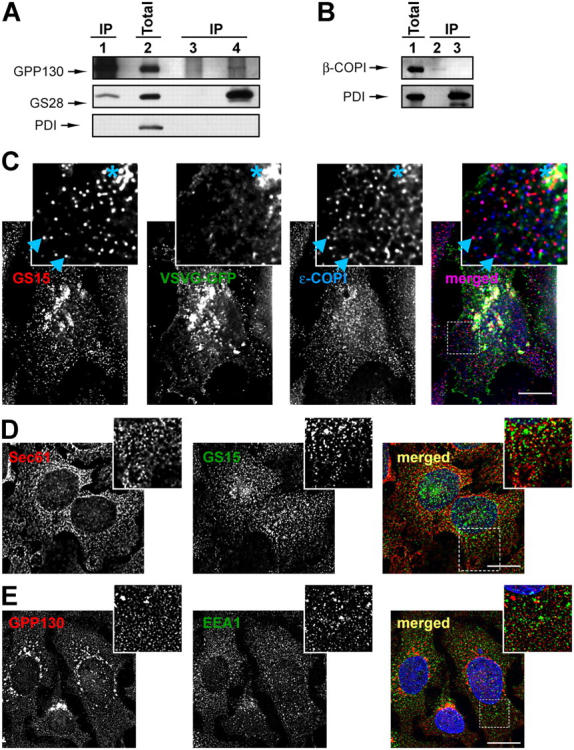
The COG complex interacts with retrograde Golgi SNARE GS28 and β-COPI. (A and B) Protein complexes from detergent-solubilized rat liver Golgi were IP using anti-Cog3p (A, lane 1; B, lane 2), anti-GS28 (A, lane 4), preimmune IgGs (A, lane 3), or anti-PDI (B, lane 3). 10% of the Golgi lysates were loaded as a control (A, lane 2; B, lane 1). Note that Cog3p, GS28, and β-COPI were not recovered with control beads or beads loaded with anti-PDI antibodies (A, lane 3; B, lane 3). (C) CCD vesicles are partially COPI coated and do not carry VSVG. COG3 KD cells that express VSVG-GFP were fixed and processed for IF with mouse anti-GS15 antibodies and rabbit anti–ɛ-COPI antibodies as described in Materials and methods. All images were acquired with 63× objective and deconvolved. Note that a number of GS15-labeled CCD vesicles were colabeled with antibodies to ɛ-COPI coat (inset, arrowheads). VSVG was partially colocalized with the GS15 only on a fragmented Golgi membrane (inset, membranes labeled with asterisk), but not on CCD vesicles. Bar, 10 μm. (D) CCD vesicles do not significantly colocalize with the ER. COG3 KD cells were fixed and stained with rabbit anti-Sec61p (red) and mouse anti-GS15 IgGs (green). Bar, 10 μm. (E) CCD vesicles are distinct from early endosomes. COG3 KD cells were fixed and stained with rabbit IgGs to GPP130 (red) and mouse anti-EEA1 IgGs (green). Bar, 10 μm.
