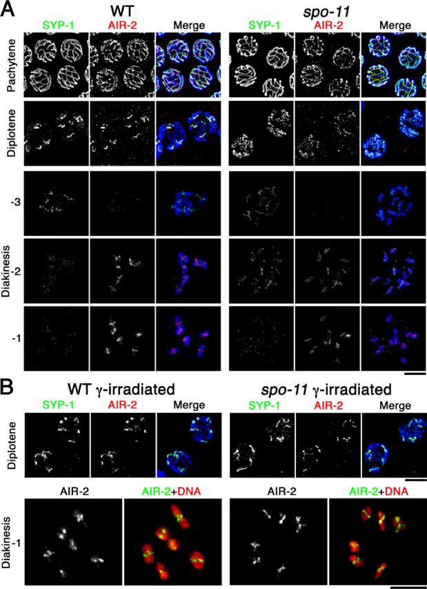
Figure 5.

Dynamic localization of SYP-1 and AIR-2. (A) Immunolocalization of SYP-1 and AIR-2 in wild-type and spo-11 nuclei at the indicated stages. For diakinesis nuclei, −3, −2, and −1 indicate position relative to the spermatheca, with the oocyte in the −1 position (closest to the spermatheca) being the most mature. In A and B, detection thresholds for α-AIR-2 signals were lower for the pachytene and diplotene panels than for the diakinesis panels to permit imaging of both the lower levels of chromosome-associated AIR-2 at the earlier stages and the subchromosomal localization of the higher levels of AIR-2 in late diakinesis. DAPI-stained chromatin is shown in blue in the merged images. (B) SYP-1 and AIR-2 localization after γ-irradiation. Bars, 5 μm.
