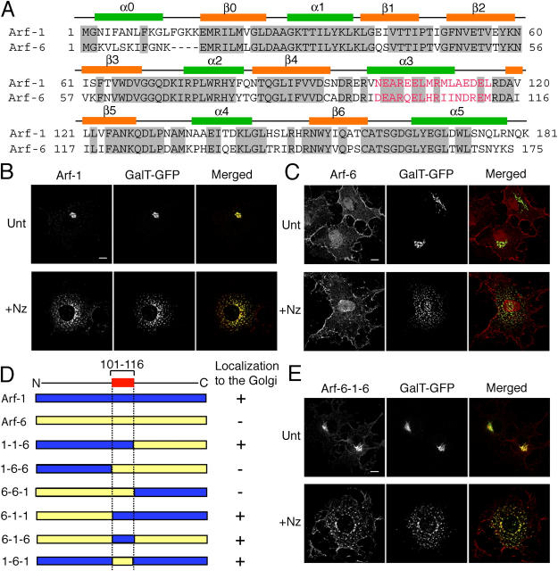
Figure 1.
Subcellular localization of Arf chimeras. (A) Numbering and secondary structural elements (α helices and β sheets) of human Arf-1 and Arf-6 are shown. Identical residues between Arf-1 and Arf-6 are shaded in gray. Residues marked in red indicate amino acids 101–116 and 97–112 of Arf-1 and Arf-6, respectively. (B, C, and E) COS-7 cells overexpressing GalT-GFP and HA-tagged Arf-1 (B), untagged Arf-6 (C), or untagged Arf-6-1-6 (E) were untreated (Unt) or incubated in the presence of 20 μg/ml nocodazole for 2 h (+Nz). Cells were fixed and immunolabeled with antibodies against HA (B) or Arf-6 (C and E) followed by Alexa 594 anti–mouse and anti–rabbit antibodies, respectively. Bars, 10 μm. (D) Diagrams of chimeras of Arf-1 and Arf-6 are shown. Blue and yellow regions represent Arf-1 and Arf-6 sequences, respectively. + and − indicate ability to localize to the Golgi.

