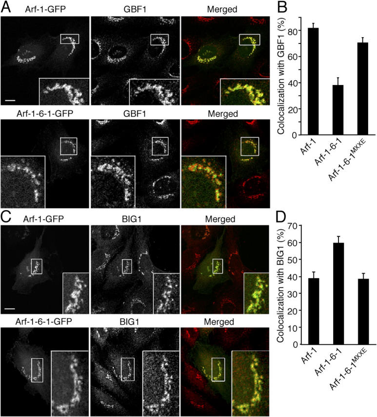
Figure 8.

Comparison of the distribution of Arf-1 and Arf-1-6-1 to Arf-GEFs. NRK cells overexpressing GFP-fused Arf-1, Arf-1-6-1, and Arf-1-6-1MXXE were fixed and immunolabeled with antibodies against GBF1 (A) or BIG1 (C) followed by Alexa 594 anti–mouse and anti–rabbit antibodies, respectively. Bars, 10 μm. (B and D) Quantitative analysis of area overlap in the Golgi region as in A and C. Error bars are the mean ± SD.
