
Figure 4.
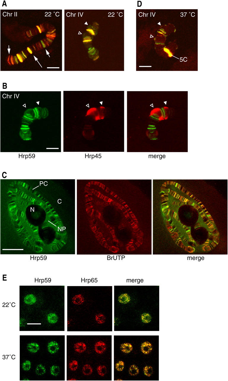
Distribution of Hrp59 and Hrp65 in the polytene chromosomes. (A) Squash preparations of C. tentans polytene chromosomes were double labeled with antibodies against Hrp59 (green) and Hrp65 (red). The BR1 and BR2 puffs in chromosome IV are indicated by arrowheads. The large arrow points to a band with intense labeling for both Hrp59 and Hrp65. The short arrows point to bands labeled by only one of the two antibodies. Note that the distributions of Hrp59 and Hrp65 are similar. (B) Polytene chromosome double labeled with antibodies against Hrp59 (green) and Hrp45 (red). The arrowheads point to the BR1 and BR2 puffs. Hrp45 and Hrp59 show different labeling patterns. (C) Transcriptionally active loci were visualized by BrUTP incorporation (red). The glands were coimmunostained with anti-Hrp59 antibody (green). C, cytoplasm; N, nucleolus; NP, nucleoplasm; PC, polytene chromosome. (D) The distributions of Hrp59 (green) and Hrp65 (red) in the polytene chromosomes were analyzed after heat shock. Note the redistribution of both proteins to the heat-shock locus at 5C (compare chromosome IV in A and D). (E) Tissue culture cells of C. tentans, either nontreated (22°C) or heat shocked (37°C), were double stained with anti-Hrp59 (green) and anti-Hrp65 (red) antibodies. All images correspond to optical sections with a thickness of ∼1 μm. Bars: (A, B, and D) ∼10 μm; (C) 20 μm; (E) 5 μm.