Figure 5.

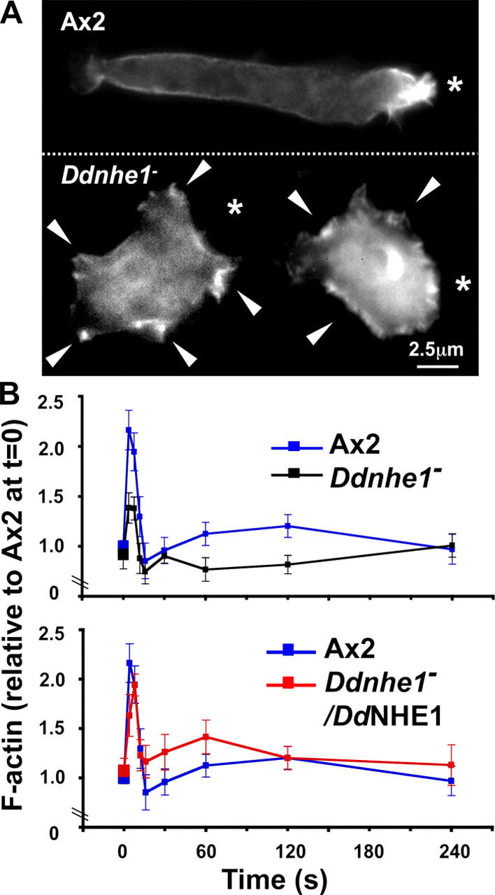
Localization and production of F-actin in response to cAMP are impaired in chemotactically competent Ddnhe1 − cells. (A) In Ax2 cells (top), F-actin was localized predominantly at the leading edge of the cell, and F-actin-rich protrusions were limited to the direction of the chemoattractant source (asterisks). In Ddnhe1 − cells (bottom), F-actin was distributed along the cell cortex, and although membrane protrusions (arrowheads) also contained F-actin, protrusions were not limited to the direction of the chemoattractant source but were seen around the cell. (B) Time course of F-actin production in response to a uniform concentration of 2 μM cAMP indicated a characteristic biphasic response in Ax2 cells (blue line), including a rapid (<10 s) first phase of greater magnitude and a slower and smaller second phase. In Ddnhe1 − cells (black line), the increase in F-actin in the first phase was reduced by ∼60% and the second phase was transient with reduced F-actin formation, compared with Ax2 cells. Ddnhe1 − /DdNHE1 cells (red line) had a biphasic increase in F-actin, although the magnitude of first and second phases was slightly greater compared with Ax2 cells. Data are expressed as the mean ± SEM of at least three independent cell preparations.
