Figure 5.

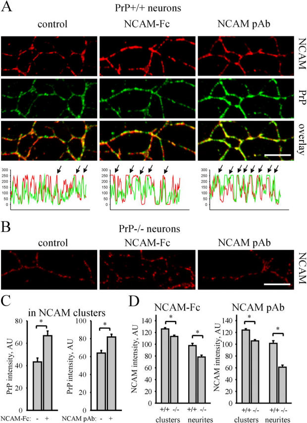
PrP is required for redistribution of NCAM to lipid rafts in response to NCAM activation. (A) PrP+/+ neurons were incubated with NCAM-Fc or polyclonal NCAM antibodies, extracted with 1% Triton X-100, and labeled with NCAM and PrP antibodies. Incubation with NCAM-Fc and NCAM pAb increased the overlap between NCAM and PrP, indicating that NCAM redistributed to lipid rafts. Examples of NCAM and PrP labeling intensities along neurites are shown below. Arrows show overlapping peaks. (B) In parallel with PrP+/+ neurons (A), PrP−/− neurons were incubated with NCAM-Fc or polyclonal NCAM antibodies (NCAM pAb), extracted with 1% Triton X-100, and labeled with NCAM antibodies. Note lower levels of detergent-insoluble NCAM in untreated (control), NCAM-Fc–, and NCAM pAb–treated PrP−/− versus PrP+/+ neurites. Bars, 10 μm (for A and B). (C) Diagrams show mean labeling intensity of PrP in NCAM clusters in control and NCAM-Fc– or NCAM pAb–incubated PrP+/+ neurons. (D) Diagrams show mean NCAM labeling intensity in NCAM clusters and along neurites of NCAM-Fc– or NCAM pAb–treated PrP+/+ and PrP−/− neurons. Mean values ± SEM (n > 50 neurites) are shown (for C and D). *, P < 0.05, t test.
