
Figure 7.
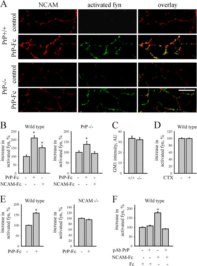
PrP-Fc activates fyn via its neuronal receptor NCAM. (A) PrP+/+ and PrP−/− neurons were incubated with PrP-Fc, extracted with 1% Triton X-100, and labeled with antibodies against NCAM and activated Tyr416 phosphorylated fyn. Note increased levels of activated fyn in NCAM clusters on PrP-Fc–treated PrP+/+ and PrP−/− neurites. Note lower levels of the activated fyn in PrP−/− neurons. Bar, 10 μm. (B) Application of PrP-Fc increased levels of activated fyn in PrP+/+ and less in PrP−/− neurons. Application of NCAM-Fc increased levels of activated fyn in PrP+/+ but not PrP−/− neurons. (C) Mean labeling intensity of GM1 along neurites of PrP+/+ and PrP−/− neurons extracted with 1% Triton X-100 is shown. (D) Aggregation of lipid rafts with cholera toxin (CTX) did not increase levels of activated fyn in PrP+/+ neurites. (E) Application of PrP-Fc increased levels of activated fyn in NCAM+/+ but not NCAM−/− neurites. (F) Application of polyclonal PrP antibodies before NCAM-Fc application blocked NCAM-Fc–induced fyn activation. (B, D–F) Diagrams show increase in mean labeling intensity of activated fyn along neurites. Mean labeling intensity of activated fyn in control neurites was taken as 100%. Mean values ± SEM (n > 50 neurites) are shown. *, P < 0.05, t test.