
Figure 5.
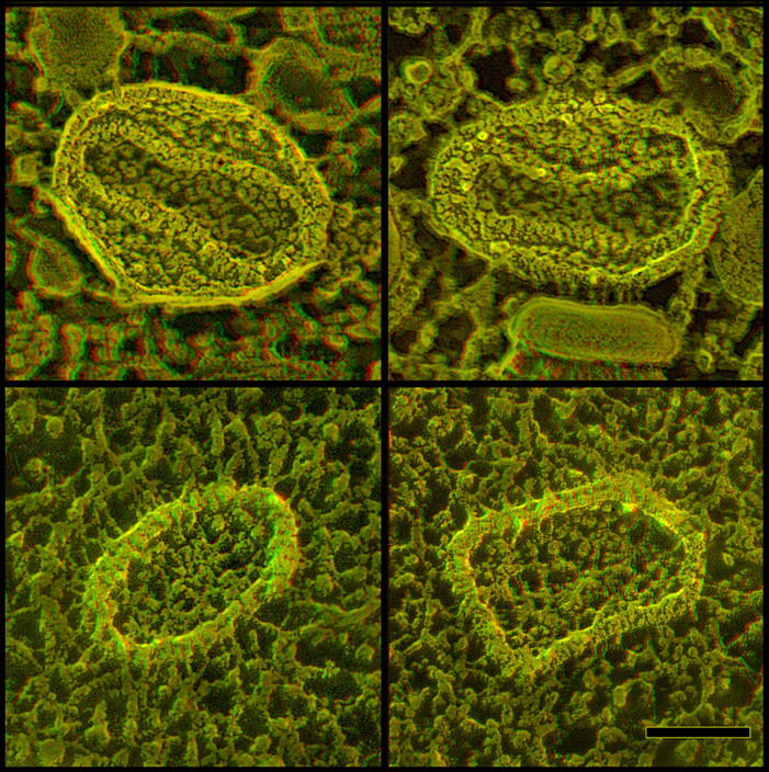
Direct comparison of the architecture and fracturing properties of the internal “palisade layer” around the cores of intact versus fused virions. (Top) Cross fractures through totally unfixed IMVs from cell like that in Fig. 2 A, showing a clear view of the internal “palisade layers” around their biconcave cores, and showing that these layers do not fracture like a biological lipid membrane. (Bottom) Crossfractures through viral cores recently released into the cytoplasm of a cell exposed to a whopping dose of vaccinia virions by “spinoculation” and quick frozen after only 5 min. The palisade layers surrounding these cores are still clearly discernible and look more or less intact. Nevertheless, even in this naked “exposed' condition in the cytoplasm, they do not freeze fracture like any membrane-containing structure. Instead, they invariably crossfracture like the palisade layers in the intact IMVs above, indicating that they are composed of nothing but protein (and polynucleotides). Bar, 0.1 μm.