
Figure 7.
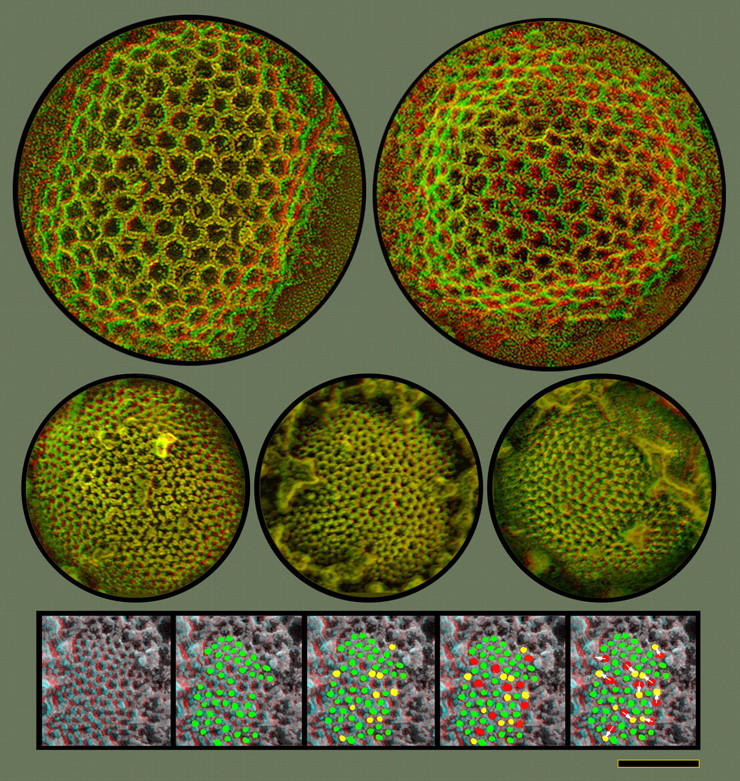
Direct comparison of clathrin lattices and the honeycomb latices on IVs, shown at exactly the same magnification. The clathrin lattices (top) have lattice parameters >2× greater than the honeycombs on IVs (center). Expansive surface views of IVs are readily obtainable because surrounding cytoplasm appears to be repelled from them, in contradistinction to IMVs (Fig. 6 A). The protein lattice observed on the IV surface is clearly a geodetic honeycomb, and sometimes shows hints of overall icosahedral symmetry. However, its lattice dimensions are much smaller. Its vertex to vertex spacing is 7 ± 1 nm versus clathrin's 15 ± 1 nm. Moreover, the vaccinia lattice typically displays many more lattice-defects and irregularities. These irregularities invariably take the form of “pentamer/heptamer” dislocations, typical for all natural honeycomb lattices. Insets at the bottom illustrate this. Proceeding from the left, green dots indicate proper hexagonal facets and yellow dots indicate inserted pentagons. Only 12 of these pentagonal insertions would be needed in the whole lattice to make it a sphere, according to Crick and Watson's postulate (Crick and Watson, 1956; Caspar and Klug, 1962); but many more pentagons are visible in this relatively small portion of the IV surface. Finally, red dots indicate inserted heptagons and white arrows indicate how each of these heptagons can be matched with a pentagon immediately adjacent to it. Bar, 0.1 μm.