
Figure 8.
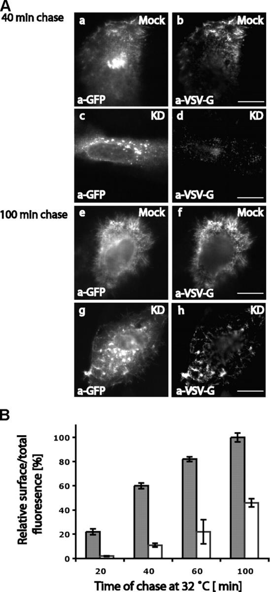
The transport of VSV-G to the cell surface is dramatically reduced in optineurin-depleted HeLa cells. Mock-treated or siRNA-treated HeLa cells were transfected with ts045-VSV-G-GFP to measure the rate of exocytosis . VSV-G at the cell surface was detected in indirect immunofluorescence using an mAb to the luminal domain of VSV-G and total VSV-G expressed was detected using a pAb to GFP. (A) Representative cells for the 40- and 100-min time points are shown to compare the amount of VSV-G on the cell surface in mock- and optineurin-depleted cells. (B) The ratio of cell surface over total VSV-G fluorescence was measured as described in Materials and methods to determine the rate of transport from the Golgi complex to the cell surface. Error bars: ±SEM. Bars, 10 μm.