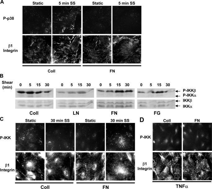
Figure 6.
Localized activation of p38 and IKK at adhesion sites. (A) Cells were plated on Coll or FN, sheared for 5 min or kept under static conditions, and stained for phosphorylated p38 and β1 integrin. Images are representative of four experiments. (B) BAE cells were plated on Coll, LN, FN, or FG and sheared for the indicated times, and IKK phosphorylation was assessed by Western blotting. Results are representative of four to six experiments. (C) Cells were plated on Coll or FN, sheared for 30 min or kept as static controls, and stained for phosphorylated IKK and β1 integrin. Results are representative of three experiments. (D) Cells on Coll or FN were treated with 10 U/ml TNFα and stained for phosphorylated IKK and β1 integrin. Images are representative of three experiments.

