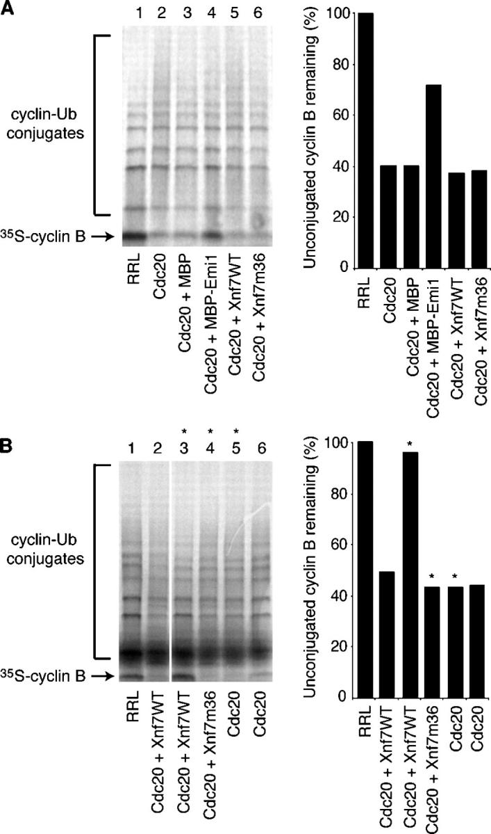
Figure 5.

Xnf7 inhibits the APC. (A) RRL (lane 1) or Cdc20 (lanes 2–6) was incubated (30 min at 23°C) with buffer (lanes 1 and 2), 6 μM MBP (lane 3), 6 μM MBP-Emi1 (lane 4), 2 μM Xnf7WT (lane 5), or 2 μM Xnf7m36 (lane 6). APC was immunopurified from mitotic egg extracts with anti-Cdc27 beads and incubated with the RRL/Cdc20 mixtures (1 h at 23°C). APC beads were washed and assayed for cyclin ubiquitylation activity using a 35S-labeled in vitro transcribed-translated Xenopus cyclin B1 fragment (1–126 aa) as a substrate. Samples were run on a 4–15% Tris-HCl SDS-PAGE gel. Unconjugated cyclin B remaining was quantitated by Phosphorimager. (B) Similar to A, except that incubations included 5 μM Xnf7WT or Xnf7m36 and were performed under conditions permissive for Xnf7's ligase activity where noted (*). Note that more than doubling the concentration of Xnf7 (compared with that used in A) did not result in APC inhibition unless the assay was performed under ubiquitylating conditions. Samples were run on a 4–20% Tris-HCl SDS-PAGE gel. White line indicates that intervening lanes have been spliced out.
