
Figure 3.
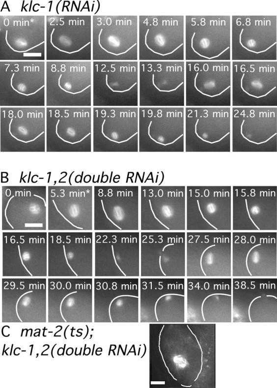
Kinesin light chains are required for normal translocation of the meiotic spindle to the cortex. Images of GFP-tubulin fluorescence are shown from representative time-lapse sequences of a meiotic embryo within a klc-1(RNAi) worm (A) and a klc-1(RNAi); klc-2(RNAi) worm (B). The cell cortex was highlighted in each image for clarity. In both cases, the meiosis I and II spindles do not move toward the cortex until after spindle shortening has initiated. Asterisks indicate exit from the spermatheca. (C) Fixed time point image of a mat-2(ts); klc-1(RNAi); klc-2(RNAi) triple mutant worm shows a meiotic spindle arrested far from the cortex. Bars, 10 μm.