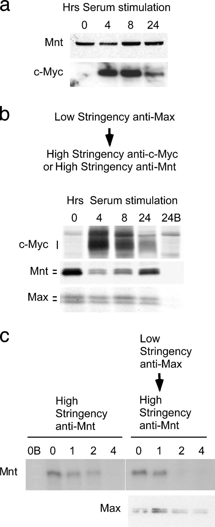
Figure 1.

Complex switching between Mnt–Max and c-Myc–Max during cell cycle entry. (a) Western blot showing Mnt and c-Myc levels at 0, 4, 8, and 24 h after serum stimulation of quiescent MEFs. (b) Levels of Mnt and c-Myc found in low stringency anti-Max immunoprecipitations during cell cycle entry. Note reduction in Mnt (and Mnt–Max complexes) at times of high c-Myc levels. (c) Pulse-chase analysis of Mnt turnover when complexed to Max. Cells were metabolically labeled with medium containing [35S]methionine (pulse) then “chased” with medium containing unlabeled methionine. Mnt was immunoprecipitated under high stringency conditions (representing total Mnt) or from low stringency Max immunoprecipitated material at 0, 1, 2, and 4 h, as indicated, during the chase period. B, immunogen blocked antibody.
