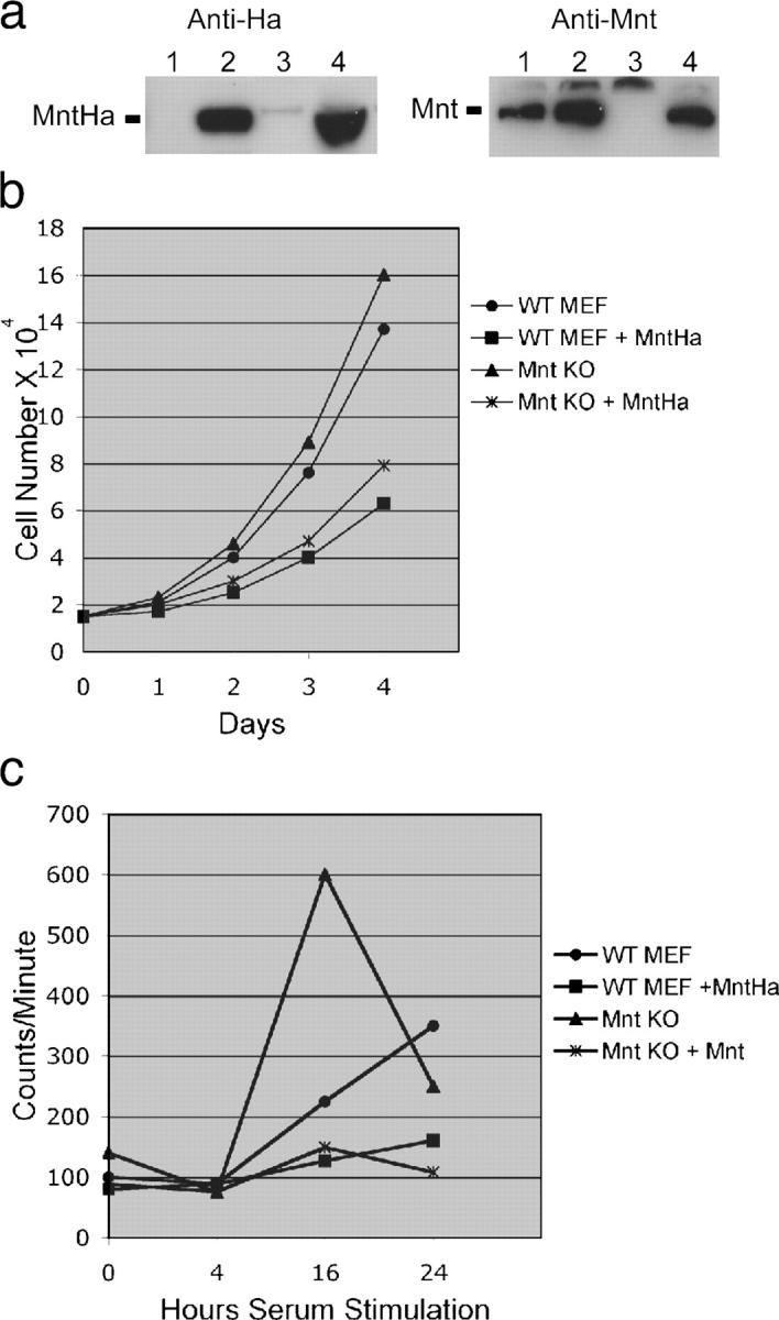
Figure 4.

Mnt overexpression slows cell proliferation and impedes cell cycle entry. (a) Western blot showing expression of Ha-tagged Mnt (anti-Ha set) in pBabeMntHa-infected MEFs (lanes 2 and 4) and endogenous Mnt (anti-Mnt set) in MEFs infected with empty virus (lanes 1 and 3). (b) Proliferation curve conducted for 4 d showing decline in proliferation rate caused by Mnt overexpression in wild-type and Mnt null MEFs. Each value is the average number of cells counted from three different dishes in two separate experiments. (c) Analysis of cell cycle (S-phase) entry in wild-type and Mnt null MEFs overexpressing Mnt. Tritiated thymidine incorporation was measured at the indicated number of hours after serum stimulation of MEFs made quiescent by confluence arrest and serum starvation. Experiments were performed at least twice in triplicate and averages are shown.
