Figure 5.

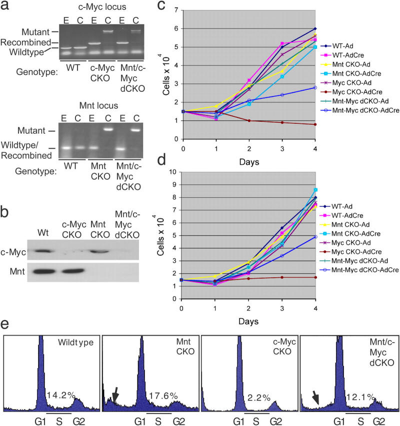
Deletion of Mnt rescues proliferation arrest caused by loss of c-Myc. (a) PCR genotyping of MEFs of the indicated genotypes after infection with empty (E) adenovirus or Cre (C) recombinase-expressing adenovirus. Note the PCR primers used to detect deleted Mnt alleles do not discriminate from wild-type and recombined Mnt alleles. (b) Western blot examining Mnt and c-Myc expression after AdCre infection of the indicated genotypes. (c and d) MEF proliferation curves after deletion of Mnt, c-Myc, and Mnt plus c-Myc in primary MEFs (c) and immortal MEFs (d). Values shown in c and d are the average of cell counts obtained from triplicate plates and are representative of results obtained from two independent experiments. (e) Cell cycle profiles generated from FAC sorting of propidium iodide stained cells 48 h after AdCre infection of the indicated cell lines. % of cells in S-phase (S) is shown. Arrows highlight sub G1 (growth phase 1) fractions consistent with apoptosis.
