
Figure 2.
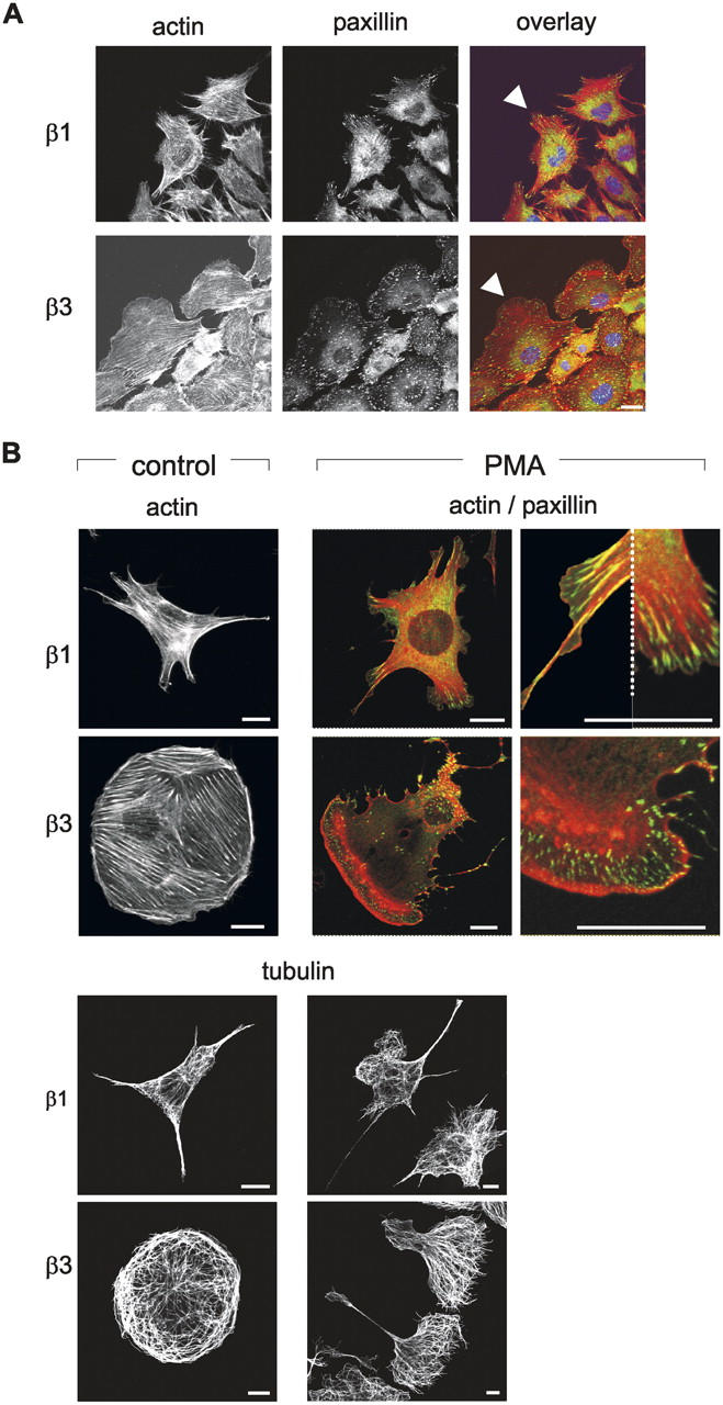
β1 and β3 integrins differentially regulate actin cytoskeletal reorganization. (A) GEβ1 or GEβ3 cells, stably expressing GFP-paxillin, were plated overnight on FN-coated coverslips, confluent monolayers were wounded with a micropipette tip, and preparations were fixed and permeabilized after 5 h. Organization of the actin cytoskeleton and localization of paxillin is shown as indicated. Arrowheads indicate protrusions of cells moving into the wounded area. Bar, 10 μm. (B) GEβ1 and GEβ3 cells were plated on FN-coated coverslips for 4 h and fixed and permeabilized either immediately (control) or after stimulation with PMA for 1 h as indicated. Single staining for F-actin, double staining for F-actin (red) and paxillin (green), or single staining for α-tubulin are shown as indicated with details of membrane protrusions shown at higher magnification at the far right. Dotted line separates two different protrusions. Bars, 5 μm.