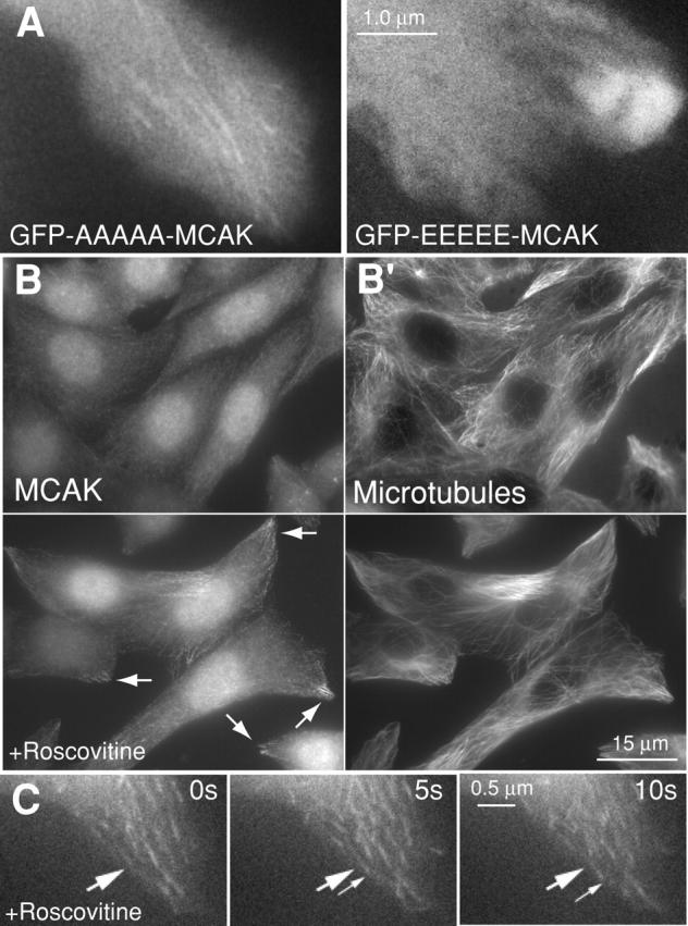
Figure 3.

Tip tracking of MCAK protein is dependent on phosphorylation. (A) GFP-AAAAA-MCAK binds to MT tips (left), whereas GFP-EEEEE-MCAK is not found on tips (right). See also Videos 4 and 5 (available at http://www.jcb.org/cgi/content/full/jcb.200411089/DC1). (B) Two fields of CHO cells labeled for endogenous MCAK (B) and MTs (B′). The top field of CHO cells are control cells. The bottom field of CHO cells were cultured for 5 h in 10 μM roscovitine and then fixed. Increased association of endogenous MCAK with distal ends of MTs is evident (B, bottom; arrows). (C) MCAK protein tracks on tips (arrows) in a HeLa cell transfected with RFP-MCAK and cultured subsequently in 10 μM roscovitine (Video 6).
