
Figure 5.
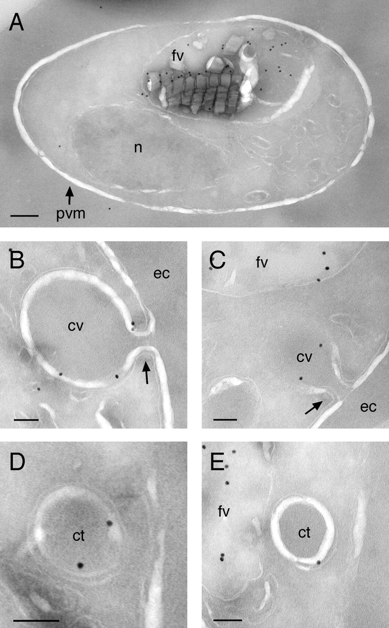
ImmunoEM localization of GFP to the food vacuole and cytostomal vacuoles. (A) A B7 trophozoite displaying prominent food vacuole labeling. Bar, 200 nm. (B and C) Cross sections of cytostomal vacuoles formed during the uptake of erythrocyte cytosol showing labeling of the vacuole membrane. In both panels, the “neck” of the cytostome (arrows) is clearly visible. (D and E) Cross sections of cytostomal vacuoles or hemoglobin transport vesicles displaying membrane labeling. Low magnification images of parasites in B–E are provided in Fig. S2. fv, food vacuole; n, nucleus; pvm, parasitophorous vacuole membrane; cv, cytostomal vacuole; ec, erythrocyte cytoplasm; ct, cytostomal vacuole or hemoglobin-containing transport vesicle. (B–E) Bars, 100 nm.