
Figure 3.
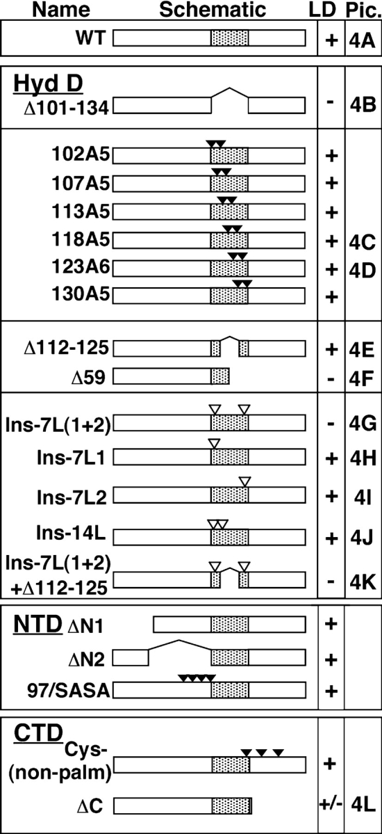
BFA-induced LD accumulation of wild-type and mutant caveolin-1. FRT cells expressing the indicated proteins were treated with BFA for 5 h, and caveolin-1 was detected by IF. Proteins are listed by name and diagrammed schematically (not to scale). The NH2-terminal, hydrophobic, and COOH-terminal domains of caveolin-1 are schematized as open, shaded, and open boxes, respectively. Deletions are schematized as gaps, substitutions as closed triangles, and 7-Leu insertions as open triangles. Except for the hydrophobic domain mutants (102A5–130A5), the number of triangles corresponds to the number of changes. Mutants in the hydrophobic domain (Hyd D), NH2-terminal domain (NTD), and COOH-terminal domain (CTD) are grouped together. Pic., IF images of these proteins are shown in the indicated panels of Fig. 4.