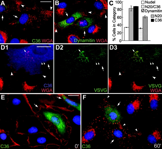
Figure 3.
Peripheral distribution of endosomes containing WGA-binding sites by Nudel mutant or dynamitin in CV1 cells. (A, B, and D–F) Large arrows indicate transfectants and concave arrowheads indicate vesicles accumulated at the cell processes. Bars, 20 μm. (A and B) Cells expressing FLAG-tagged NudelC36 or dynamitin were fixed in methanol and labeled with TRITC-WGA (red), anti-FLAG mAb (green), and DAPI (blue). (C) Statistic results (mean ± SD) showing severity of vesicle dispersion. n = 300, three experiments. (D) Distributions of VSVG-GFP (green) and WGA-positive vesicles (red) in a typical FLAG-NudelC36 expressor (blue). Small arrows and arrowheads indicate the Golgi cisternae and typical secretory vesicles, respectively. (E and F) Binding of WGA (red) to the plasma membrane at 4°C and its endocytosis into GFP-NudelC36 expressors (green) after 60 min at 37°C.

