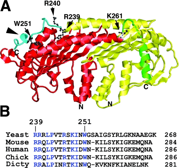
Figure 1.
A model structure for yeast CP. (A) Cap1 and Cap2 are colored yellow and red, respectively, except that their proposed tentacles are cyan and green, respectively. Certain residues targeted for mutation are indicated. Cap2 residues 268–273 could not be modeled and are indicated as a dashed line. Arrowhead indicates amphipathic α-helix in the COOH-terminal region of the α subunit. (B) Alignment of the COOH-terminal regions of CP α in various species. Residues found in all species are colored blue.

