Figure 6.

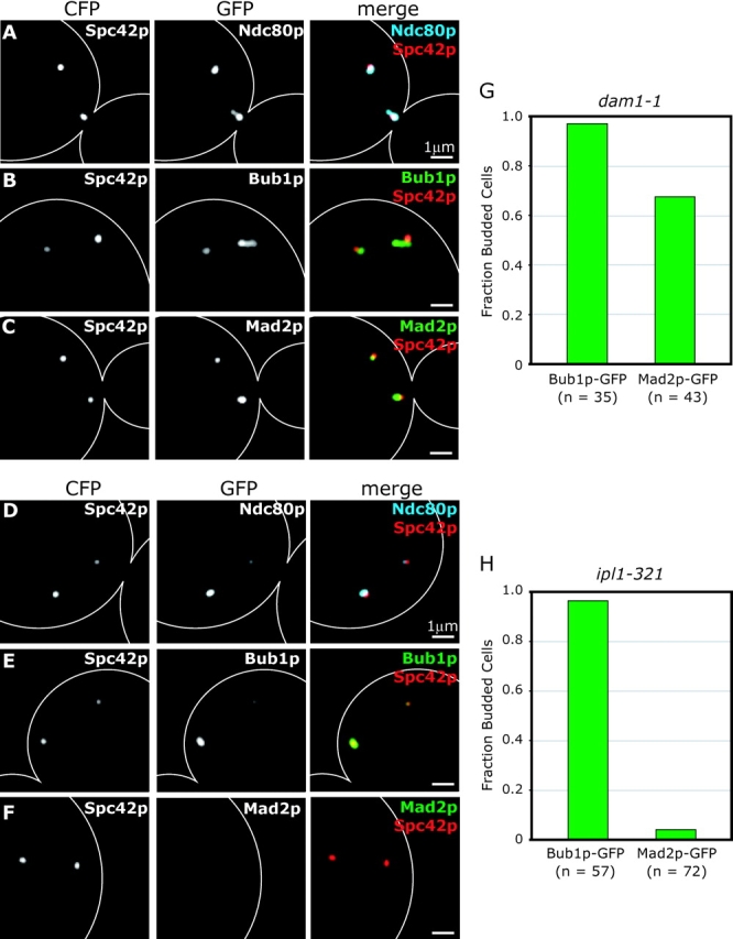
Bub1p and Mad2p localization in dam1-1 and ipl1-321 cells. (A–C) dam1-1 cells expressing the SPB protein Spc42p-CFP and Ndc80p-GFP, Bub1p-GFP or Mad2p-GFP at nonpermissive temperature. Cells were grown at 25°C to mid-log phase and shifted to 37°C for 1 h before fixation. Panels show Spc42p-CFP alone; Ndc80p-GFP, Bub1p-GFP, or Mad2p-GFP; and Spc42p-CFP (red) merged with Ndc80p-CFP (blue), Bub1p-GFP (green), or Mad2p-GFP (green). (D–F) ipl1-321 cells expressing Spc42p-CFP and Ndc80p-GFP, Bub1p-GFP, or Mad2p-GFP at nonpermissive temperature. Cells were arrested in α-factor for 2 h, shifted to 37°C for 10 min and released at 37°C for 2 h before fixation. (G and H) Fraction of budded cells containing Bub1p-GFP and Mad2p-GFP for dam1-1 cells after 1 h at 37°C and ipl1-321 cells after 2 h at 37°C after α-factor release. n = number of budded cells counted.
