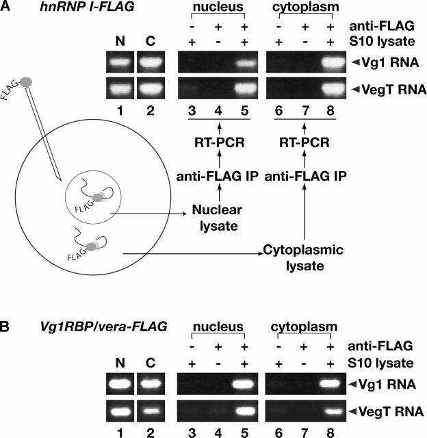
Figure 2.
hnRNP I and Vg1RBP/vera associate with Vg1 and VegT RNAs in the nucleus and cytoplasm. (A) Stage III/IV oocytes were injected with RNA encoding hnRNP I-FLAG, and S10 lysates were prepared from manually isolated nuclei and cytoplasm. Vg1 (top) or VegT (bottom) RNAs in the nucleus (lane 1) or cytoplasm (lane 2) were detected using RT-PCR. Immunoprecipitations were performed using S10 lysates with Sepharose beads (lanes 3 and 6), anti-FLAG beads in the absence of lysate (lanes 4 and 7), and anti-FLAG beads in the presence of nuclear S10 (lane 5) or cytoplasmic S10 (lane 8) lysate. Vg1 and VegT RNAs were detected in each immunoprecipitate by RT-PCR. All samples were run on the same gel, but lane order was changed for presentation in the figure. (B) Stage III/IV oocytes were injected with RNA encoding Vg1RBP/vera-FLAG and S10 lysates were prepared as in A. Vg1 (top) or VegT (bottom) RNAs in the nucleus (lane 1) or cytoplasm (lane 2) were detected by RT-PCR. Immunoprecipitations were performed using S10 lysates with Sepharose beads (lanes 3 and 6), anti-FLAG beads in the absence of lysate (lanes 4 and 7), and anti-FLAG beads in the presence of nuclear (lane 5) or cytoplasmic (lane 8) S10 lysate. Vg1 RNA and VegT RNA were detected in each immunoprecipitate by RT-PCR; all samples were separated on the same gel, but lane order was changed for presentation in the figure.

