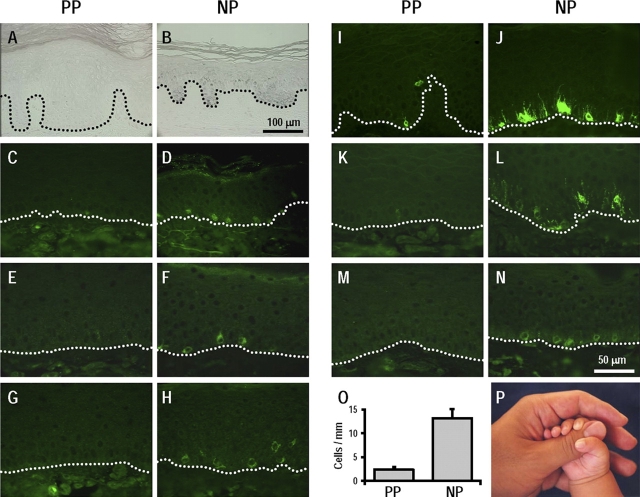
Figure 1.
Melanocyte function in palmoplantar (PP) and in nonpalmoplantar (NP) skin. (A and B) Fontana-Masson staining for melanin. Bar, 100 μm. (C–N) Immunohistochemical staining for MITF (C and D), TYR (E and F), DCT (G and H), MART1 (I and J), and gp100 (K–N). HMB45 (K and L) and αPEP13h (M and N) specifically stain gp100 in stage II–IV melanosomes and in stage I melanosomes, respectively. Bar, 50 μm. (O) Melanocyte density measured by the number of cells positive for melanosomal proteins. Data are reported as means ± SD. (P) Macroscopic view of hypopigmented palm (palmoplantar) skin and hyperpigmented arm (nonpalmoplantar) skin.

